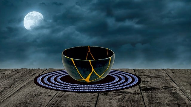
Figure 1: Image showing that protein-based artificial kinetochores (magenta) can interact with spindle microtubules (green) in a similar manner to chromosomes (cyan). © 2026 Yuanzhuo Zhou
For sexual reproduction to yield healthy offspring, newly generated oocytes-immature egg cells-must receive the correct amount of DNA after cell division. This process of segregating chromosomes becomes more prone to failure as we age. Now, RIKEN researchers have identified a strategy that could help to prevent such errors and restore healthy production of oocytes1.
Oocytes are produced by a cell-division process known as meiosis, during which every chromosome is duplicated. These replicates form X-shaped structures in which the chromosomes are joined via structures called centromeres, where a protein called cohesin locks chromosome copies together.
As division proceeds, protein fibers called microtubules spread from opposite poles of the dividing cell, attaching to each chromosome. These microtubules eventually pull the two apart, so that each newly formed cell receives one copy of each chromosome.
However, older mothers have less cohesin. "Studies using mouse models show that the amount of cohesin complexes gradually declines on oocyte chromosomes with age," explains Tomoya Kitajima of the RIKEN Center for Biosystems Dynamics Research. "Similar issues are likely to arise in aging human oocytes as well."
This means coupled chromosomes may separate prematurely, leading to abnormal distribution of genomic material and yielding ova that may exhibit developmental defects or fail to produce viable offspring.
Kitajima was initially interested in understanding this breakdown in cell division, but now he may have found a way to remedy it.
"This project originally began with a motivation to build an artificial kinetochore," he says, referring to the chromosomal structures that microtubules lock onto during meiosis.
But as his team began dabbling with these constructs in dividing mouse oocytes, they realized that these artificial kinetochores could compete with actual chromosomal kinetochores for microtubule binding. This reduced the overall pulling force applied to chromosome pairs, such that even those with weakened cohesion remained coupled.
They have now demonstrated their latest-generation artificial kinetochores, formed from compact protein assemblies that can be directly expressed in oocytes.
Meiosis is a two-stage cell division process, ultimately yielding gametes with just one of each chromosome rather than the two copies found in normal cells. The team's artificial kinetochores helped normalize distribution of chromosomal DNA at both stages in aged mouse cells.
This could one day lead to treatments that safeguard healthy conception in older parents. "This strategy is far from ready for clinical application," cautions Kitajima. "But I believe it could become feasible in the future."
To advance towards this goal, he aims to develop animal models that more faithfully capture the complexities of human production of oocytes as a testbed for their artificial kinetochore strategy.