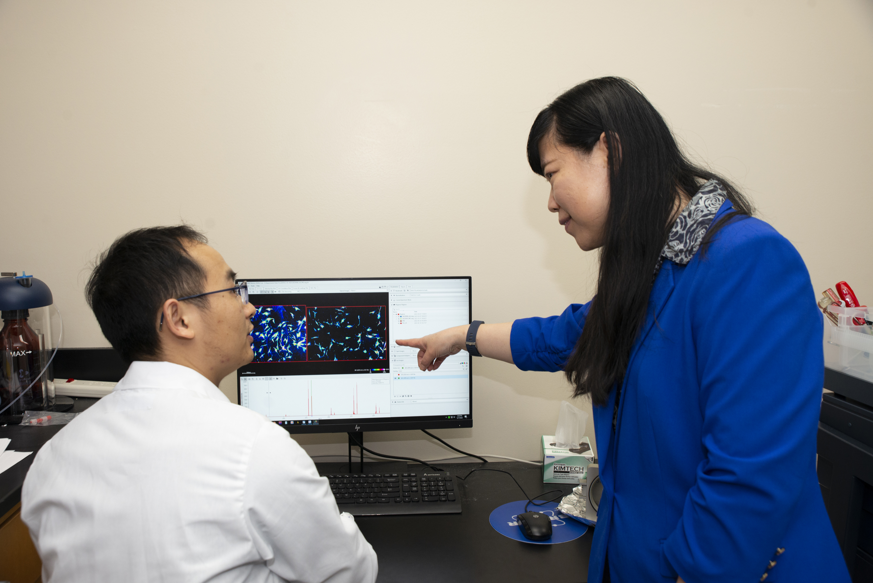
Scientist Hua Zhang, left, and UW-Madison professor Lingjun Li analyze a mass spectrometry image in Li's lab in the School of Pharmacy.
Research at the University of Wisconsin-Madison drives innovation, saves lives, creates jobs, supports small businesses, and fuels the industries that keep America competitive and secure. It makes the U.S.-and Wisconsin-stronger. Federal funding for research is a high-return investment that's worth fighting for. Learn more about the impact of UW-Madison's federally funded research and how you can help.
Lingjun Li, a professor in the University of Wisconsin-Madison School of Pharmacy and Department of Chemistry, has spent decades developing powerful new ways to measure and map the molecular machinery of life.
Recently, Li and her collaborators at UW-Madison and the Howard Hughes Medical Institute introduced a new imaging technology that has the power to reveal biomolecular detail in tissues like cancer tumors in their native environments and at unprecedented resolution. That technology, recently reported in Nature Methods, has been described as a potential game-changer for biomedical researchers.
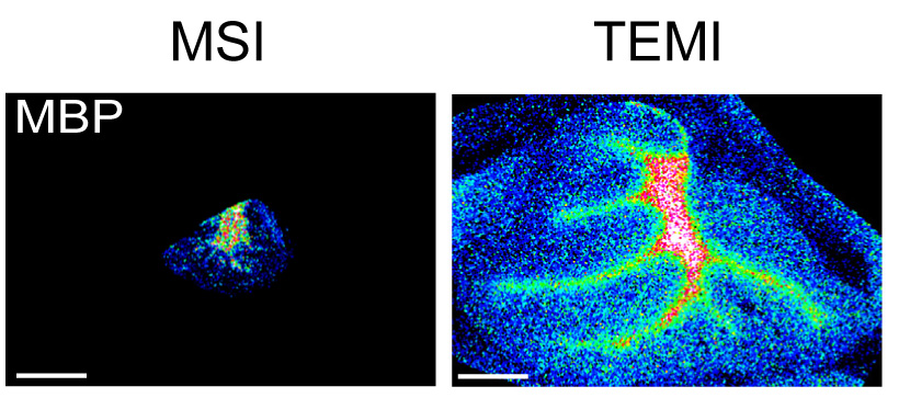
This image demonstrates the difference between standard mass spectrometry imaging (left) and the new technology developed by Li and her colleagues (right), which provides significantly more detail. The images show a peptide from a mouse cerebellum.
Li's track record for advancing mass spectrometry and other techniques earned her a spot on The Analytical Scientist's 2024 Power List, and she recently received a Kellett Mid-Career Award from the UW-Madison Office of the Vice Chancellor for Research. The award honors mid-career tenured faculty who have made key research contributions in their field.
Here, Li reflects on her lab's interdisciplinary research, the importance of spatial molecular imaging, and how tools like AI and mass spectrometry are shaping the future of biomedical science.
Can you describe your research background and what brought you to UW-Madison?
I'm a bioanalytical chemist by training - my background is in analytical and biomolecular chemistry. I did a unique postdoc, spending a year at Pacific Northwest National Lab working on high-end mass spectrometry-based proteomics, and another year at Brandeis University in a neurobiology lab using crabs and lobsters as model organisms. I joined UW-Madison in December 2002 to combine mass spectrometry technology development with fundamental neuroscience research, always with the goal of improving human health.
How has your lab's focus evolved since then?
Initially, we aimed to discover nervous system biomolecules called neuropeptides and understand their function. Over time, we've expanded to studying a wide variety of biomolecules - proteins, peptides, lipids, metabolites, glycans - and their spatial distributions in tissues. Mass spectrometry allows us to look at all of these molecules with high specificity. A major focus now is understanding how these molecules contribute to diseases like Alzheimer's, work that we're doing in collaboration with the Wisconsin Alzheimer's Disease Research Center.
What makes mass spectrometry a powerful tool for your work?
Mass spectrometry measures the mass of molecules and can provide structural information about them. For proteins, we can determine amino acid sequences and their modifications. For lipids and metabolites, we can distinguish structural isomers - molecules with the same mass but different chemical bonds. That structural detail can be crucial, for example, in distinguishing cancer cells from healthy cells.
What's the significance of spatial context in molecular imaging?
Cells, even adjacent neurons, can use different chemical messengers. Without spatial information, you lose context about how molecules function in specific areas. In cancer, for instance, being able to see tumor heterogeneity at a fine level could influence treatment strategies. That's why we're focused on imaging techniques that retain spatial resolution.
Your lab recently helped develop a new mass spectrometry imaging technique that's been described as a game-changer. What's new about it?
We integrated tissue expansion microscopy with mass spectrometry imaging. Traditional imaging mass spectrometry lacks spatial resolution and loses the important context of how molecules behave in their natural environment within tissue. By physically expanding the tissue under mild conditions, we preserve its molecular composition and native structure while achieving higher resolution without needing fancy or expensive new hardware. That makes this approach, led by Hua Zhang in my lab, both powerful and accessible.
Why is making this new technique accessible important to you?
We want this to be open and available to biomedical researchers everywhere. Anyone with a commercial mass spectrometer can use the technique, and tissue expansion itself follows a straightforward protocol. What's exciting is that you can achieve higher resolution without expensive new equipment or long acquisition times. That opens up mass spectrometry imaging to more biologists - helping them investigate molecular detail down to the single-cell level.
How do you see analytical science contributing to broader research and societal needs?
Analytical science is central to so many disciplines. It's not just about supporting other research - it's a science of measurement. Whether it's environmental pollutants like PFAS or biomarkers in human disease, we need tools that are sensitive and precise. Mass spectrometry-based approaches are increasingly seen as foundational tools in biological discovery. Analytical science enables system-level investigations that not only uncover new biological mechanisms but also inspire the development of innovative hypotheses and transformative technologies.
How is your lab thinking about AI and machine learning in this space?
We're generating huge volumes of data. Machine learning can help us translate these data into biological and clinical insights. We already use clustering and algorithmic tools for things like neuropeptide identification and single-cell analysis. Collaborating with statisticians and developing new software is key to managing this complexity. AI has great potential for biomarker discovery and predictive modeling in precision medicine.
What makes your lab environment unique?
We're very interdisciplinary. My group includes students and postdocs from analytical chemistry, pharmaceutical sciences and biophysics. That diversity helps us tackle big, meaningful problems. And analytical science is very practical - it not only advances health-related research but also prepares our trainees for real-world careers in academia, industry and public health.
What motivates your continued focus on technology development?
Our goal is always to develop tools that can improve human health. We don't build technology for its own sake - it's always about enabling discovery. Whether it's understanding disease mechanisms, identifying early biomarkers, or informing treatment strategies, analytical science has the potential to make a real difference.
The research that led to the new technology recently described in Nature Methods was supported in part by the National Institutes of Health (grants R01AG078794, R01DK071801, R01AG052324, P01CA250972 and DP1DK113644) and the U.S. Department of Agriculture (2018-67001-28266).


