By Joey Garcia, University Communications and Marketing
It infects nearly one-third of the global population, yet its microscopic size makes the parasite difficult for scientists to study.
That parasite is Toxoplasma gondii, a widespread organism that infects humans and animals. To better understand how it functions, infectious disease researchers at the USF Health Morsani College of Medicine adapted a fluorescent imaging system typically used to study human cells to observe the parasite's growth in real time-paving the way for future treatments.
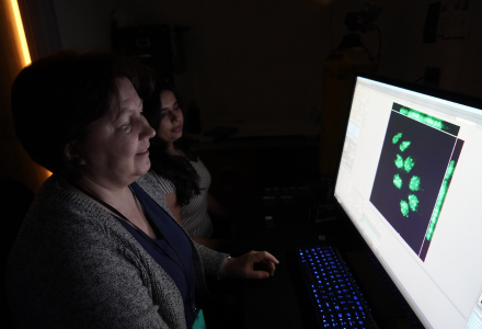
Elena Suvorova and Mrinalini Batra observing the detailed toxomplasma cells
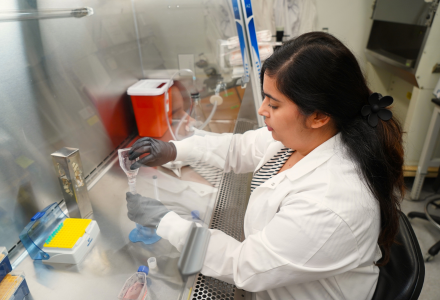
Mrinalini Batra prepares the toxomplasma gondii parasite for observation in the Suvorova Lab
"Our adapted technology will now give scientists the first clear and instant view of how this parasite grows. With that level of detail, we can better understand its biology and identify where it may be vulnerable for better interventions."
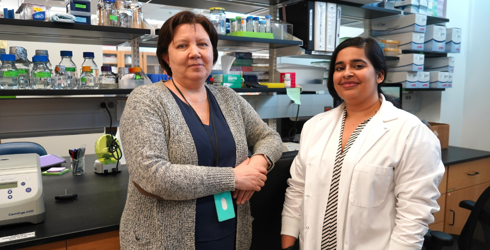
USF Associate Professor Elena Suvorova
AN UNUSUAL CELL CYCLE
Toxoplasma is commonly spread through uncooked meat and contaminated produce. When the parasite enters the human body, it causes toxoplasmosis, an infection that is often mild, but can be serious for pregnant women and people with weakened immune systems. It can be treated if caught within the first two weeks of exposure.
"Though the parasite can be repressed in the acute stage, it requires drugs that can be toxic if taken long term," Suvorova said. "If you can't catch toxoplasmosis during this time, the parasite turns chronic. In this stage, it hides from the immune system and forms cysts in the brain, for which there are currently no cures."
Developing improved treatments has been challenging in part because of the parasite's unusual cell cycle. A typical cell cycle begins with the cell growing larger before making a complete copy of its DNA. Once everything is prepared for division, the cell splits into two identical parts.
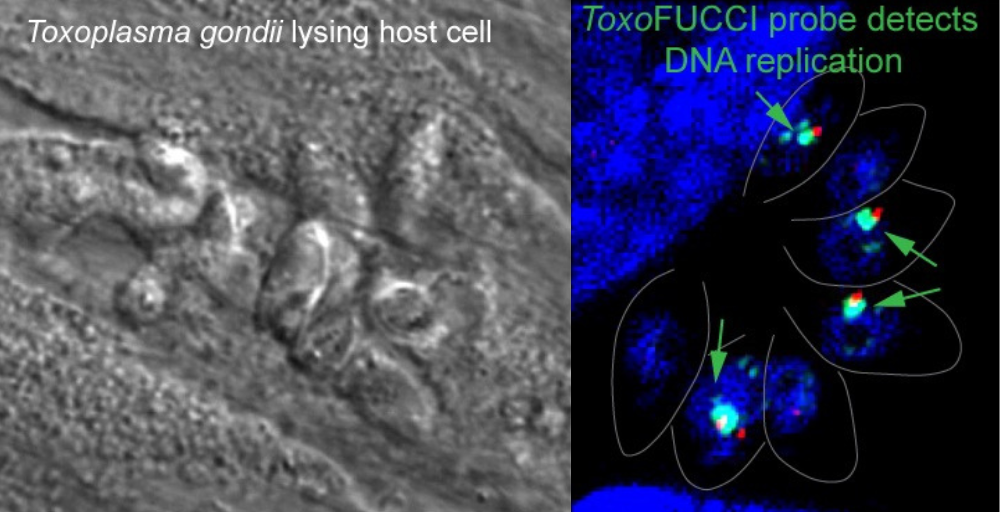
Fluorescent imaging allows scientists to observe the parasite Toxoplasma gondii in far greater detail, revealing how it replicates and progresses through its cell cycle
"Toxoplasma doesn't follow this standard pattern," said Mrinalini Batra, a research scientist in the Suvorova Lab."Scientists knew it had to go through similar stages because it reproduces, but they didn't know how those stages were arranged or whether they even existed in the same way as they do in human cells. That made it hard to understand how this parasite grows and spreads."
The goal of the researchers wasn't just basic curiosity, but part of a bigger effort to eventually stop the parasite from multiplying. To do that, the team needed to map out how its cell cycle works and in what order.
TARGETING TOXOPLASMA THROUGH FLUORESCENCE
To tailor their fluorescent imaging model for Toxoplasma, the team first had to identify proteins that appear in specific growth stages of the parasite. These proteins also needed to be located in structures that are large enough to visualize, such as the cell's nucleus, and required fluorescent colors bright enough to stand out in such a tiny organism under a microscope.
Because Toxoplasma lacks many of the common proteins found in human cells, the process required extensive trial and error. The team tested different parts of the parasite using red and green fluorescent tags, but many markers either failed to glow brightly enough or did not appear in sufficient amounts to be useful.
As the team tested multiple combinations, they ultimately identified a protein called PCNA1. This protein is located in the parasite's nucleus and naturally shifts as the organism progresses through its growth cycle.
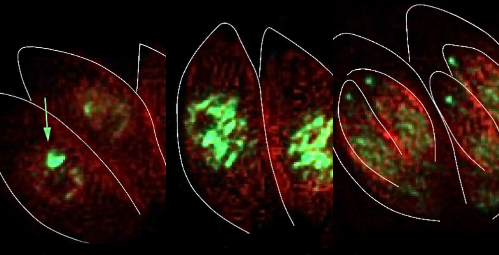
Fluorescent tagging of the PCNA1 protein (green) reveals when the parasite Toxoplasma gondii begins copying its DNA, allowing researchers to track key stages of its cell cycle in real time
"When we attached two copies of a bright neon green tag to this protein, the signal became strong and clear," Batra said. "This allowed us to determine the parasite's stage simply by watching how the glowing protein behaved in the cell cycle. For the first time, researchers were able to clearly map Toxoplasma's cell cycle."
The discovery shows how the parastate moves normally through the first part of its cell cycle, but the rest of its growth stages overlap instead of occurring sequentially.
The Suvorova Lab plans to continue its work, analyzing the parasite to identify opportunities for targeted intervention early in its development
"These latter stages are similar to a fork's structure," Suvorova said. "Toxoplasma's cell cycle begins with one straight handle and then several prongs that branch off, allowing as many as three cell cycle phases to occur simultaneously. This unusual pattern helps the parasite multiply rapidly and evade the host's immune system before forming cysts in the brain."
Now that Toxoplasma's cell cycle has been mapped through fluorescence, the team is working to identify weak points in the parasite that could prevent it from multiplying. The team is also testing how different drugs affect specific stages of the cycle in hopes of developing safer and more effective treatments.


