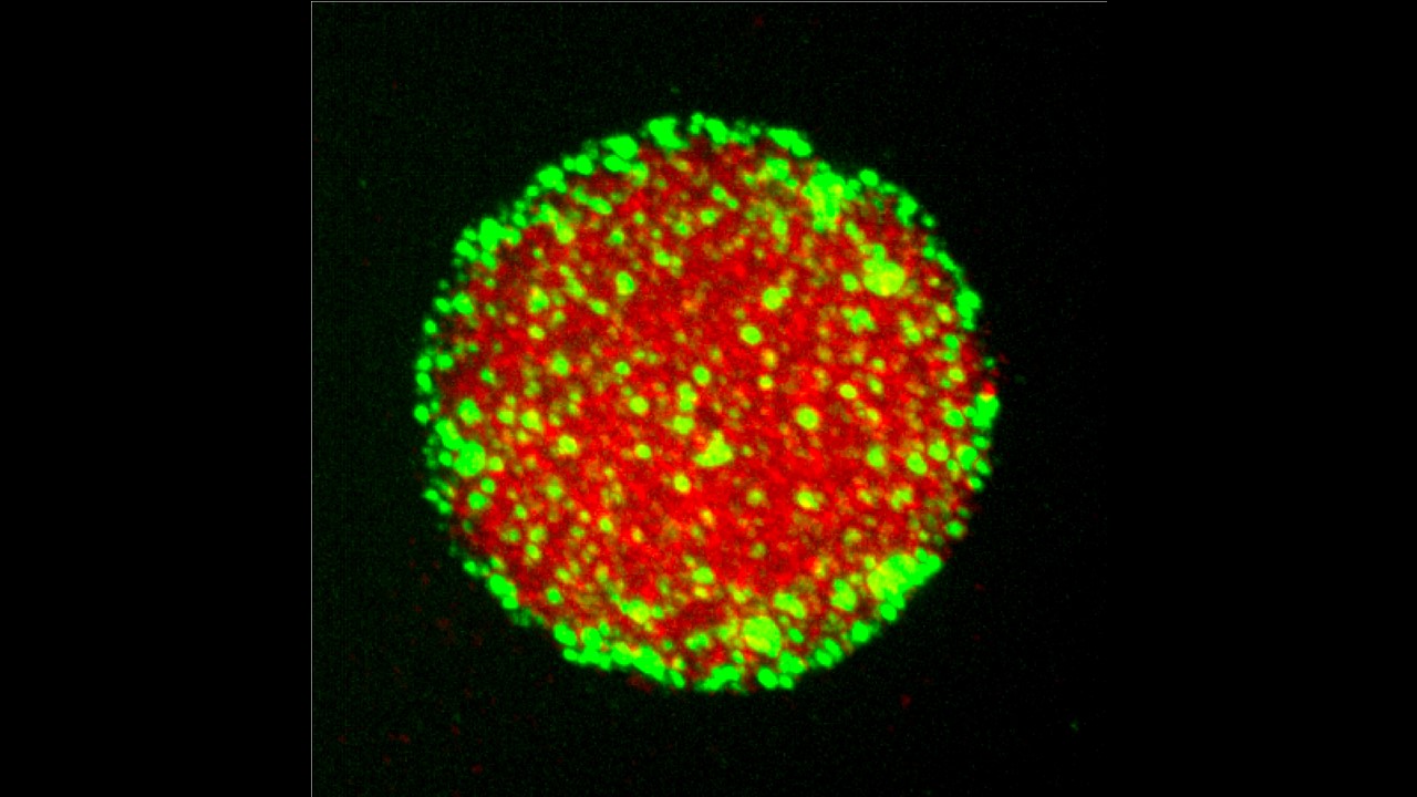
Protein condensates (red) are stabilized by Pickering agents (green) that adsorb to the condensate surface. Credit: Andrew Folkmann
Johns Hopkins Medicine researchers report that food science principles have helped them determine how unusual droplets within cells stay organized and avoid dissolving into the rest of the cell's gelatinous interior.
The researchers say their work could advance scientific understanding of cell evolution and help scientists in the food and chemical industry develop better ways to keep liquid mixtures from separating.
The cells of all living organisms hold a collection of mini biological machines called organelles. These structures run the cell's powerhouse mitochondria, brainy nucleus and other operations, all with a defined border and encased in a membrane. However, there are other cell parts that appear as viscous, membrane-free "blobs," but they serve distinct purposes, such as regulating genes, sending chemical signals or storage sites for specialized molecules.
Scientists have long thought these somewhat mystifying droplets might be a primordial version of organelles, and the Johns Hopkins-led research team worked with laboratory worms to study them further.
A report on the research team's findings about these droplets, which are called biomolecular condensates, appears Sept. 10 in Science.
"I hope this work will help convince scientists that biomolecular condensates are highly sophisticated cellular compartments," says Geraldine Seydoux, Ph.D., the Huntington Sheldon Professor in Medical Discovery and vice dean for basic research at the Johns Hopkins University School of Medicine and investigator at the Howard Hughes Medical Institute. "We found they have regulated roles and respond to the environment, just like other organelles. And we found that they do have membranes, just not the type we're used to seeing."
Biomolecular condensates were first dubbed "granules" in the 1970s by scientists who used electron microscopy to peer more closely at the structures in many organisms, including squiggly creatures called C. elegans, whose relatively simple biology has made them a common laboratory model for studying everything from modern gene-cutting technology to protein structure. The condensates in worms, which look tough and similar in appearance to grains of sand, are known as P granules.
In 2014 in Seydoux's lab, graduate student Jennifer Wang conducted genetic analyses to find a protein called MEG-3 in worm P granules. Wang's experiments showed that another protein, PGL-3, creates the viscous liquid droplets, the "core" of P granules, and that MEG-3 loiters on the outside of the P granule, making small "clusters" that coat the surface of the P granules.
"What we didn't understand was these proteins could just linger on the outside of P granules yet be so integral to stabilizing the interior of the granules," says Seydoux.


