UNIGE scientists have reconstructed for the first time a film of the assembly of the human centriole, one of the essential structures that constitute our cells.
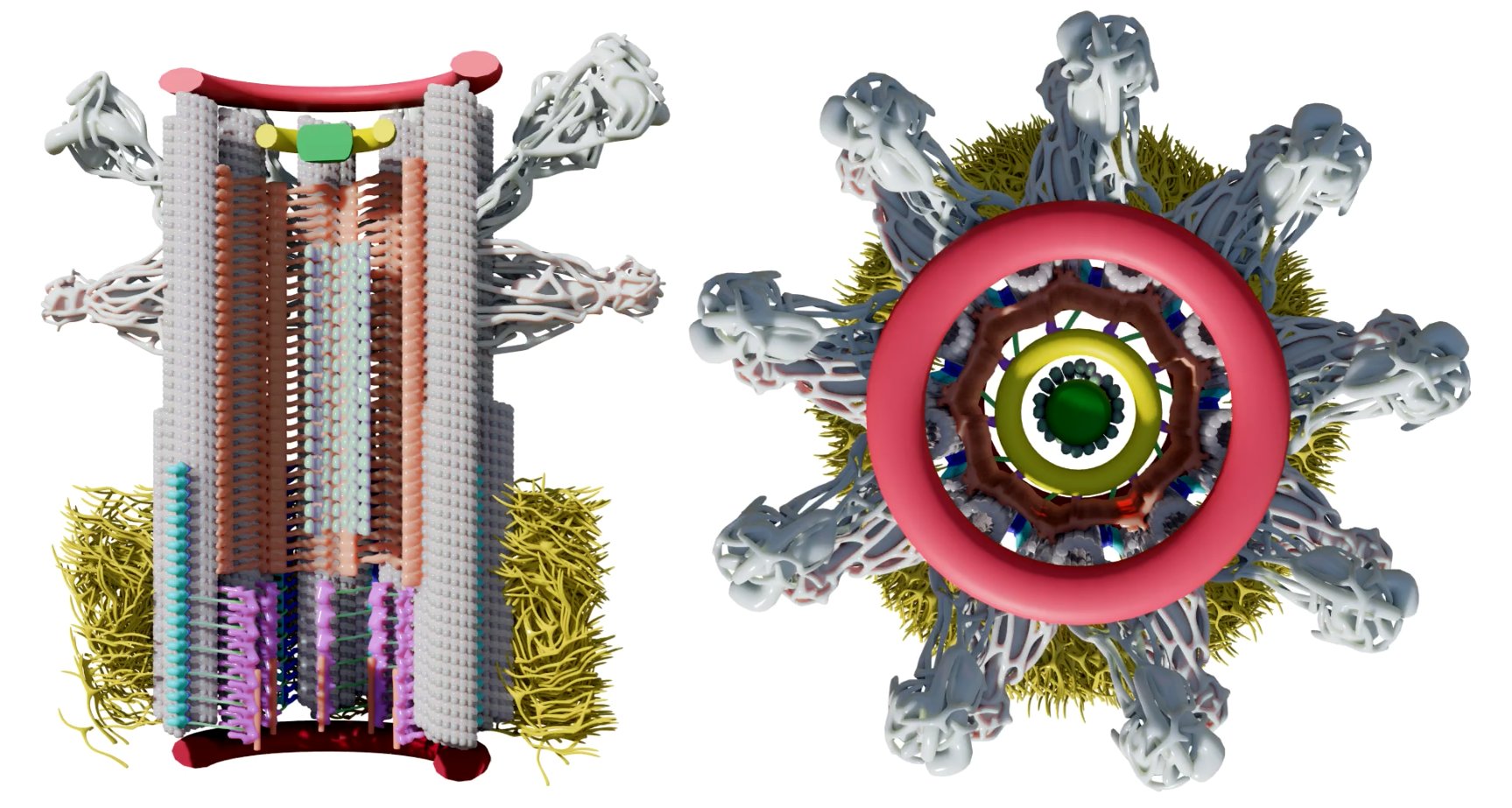
Model of a human centriole cut along its longitudinal axis and viewed from above. © CentrioleLab
Cells contain various specialised structures - such as the nucleus, mitochondria or peroxisomes - known as "organelles''. Tracing their genesis and determining their structure is fundamental to understanding cell function and the pathologies linked to their dysfunction. Scientists at the University of Geneva (UNIGE) have combined high resolution microscopy and kinematic reconstruction techniques to visualise, in motion, the genesis of the human centriole. This organelle, essential to the organisation of the cell skeleton, is associated - in case of dysfunction - with certain cancers, brain disorders or retinal diseases. This work, published in the journal Cell, elucidates the complexities of centriole assembly. It also opens up many new avenues for the study of other cell organelles.
Organelle genesis proceeds according to a precise sequence of successive protein recruitment events. Visualising this assembly in real time provides a better understanding of the role of these proteins in organelle structure or function. However, obtaining a video sequence with sufficient resolution to distinguish such complex microscopic components faces a number of technical limitations.
Inflating cells for better observation
This is particularly true of the centriole. This organelle, measuring less than 500 nanometers (half a thousandth of a millimeter), is constituted of around 100 different proteins organised into six substructural domains. Until a few years ago, it was impossible to visualise the structure of the centriole in detail. The laboratory of Paul Guichard and Virginie Hamel, co-directors of research in the Department of Molecular and Cellular Biology at the UNIGE Faculty of Science, has changed this situation by using the technique of expansion microscopy. This cutting-edge technique enables cells and their constituents to be progressively inflated without being deformed, so that they can then be observed - using conventional microscopes - with very high resolution.
"We were able to put these thousands of images taken at random back into chronological order, to reconstruct the various stages in the formation of centriole substructures."
Obtaining images of the centriole with such high resolution enables the exact location of proteins at a given time but gives no information on the order of appearance of substructural domains or of individual proteins. Marine Laporte, a former research and teaching fellow in the UNIGE group and first author of the study, used expansion microscopy to analyse the location of 24 proteins in the six domains in over a thousand centrioles at different stages of growth.
Reorganising images to set them in motion
''This very tedious work was followed by a pseudo-temporal kinematic reconstruction. In other words, we were able to put these thousands of images taken at random during centriole biogenesis back into chronological order, to reconstruct the various stages in the formation of centriole substructures, using a computer analysis we developed,'' explains Virginie Hamel, co-leader of the study.
This unique approach, which combines the very high resolution of expansion microscopy and kinematic reconstruction, has enabled us to model the first 4D assembly of the human centriole. ''Our work will not only deepen our understanding of centriole formation, but also open up incredible prospects in cellular and molecular biology, since this method can be applied to other macromolecules and cellular structures to study their assembly in space and time,'' concludes Paul Guichard.


