Key Points
- Griffith University researchers investigated dental implant healing by focusing on the role of osteocyte lacunae in osseointegration
The study provides knowledge to help improve the design of implant surfaces and the selection of materials used in implants to improve patient healing
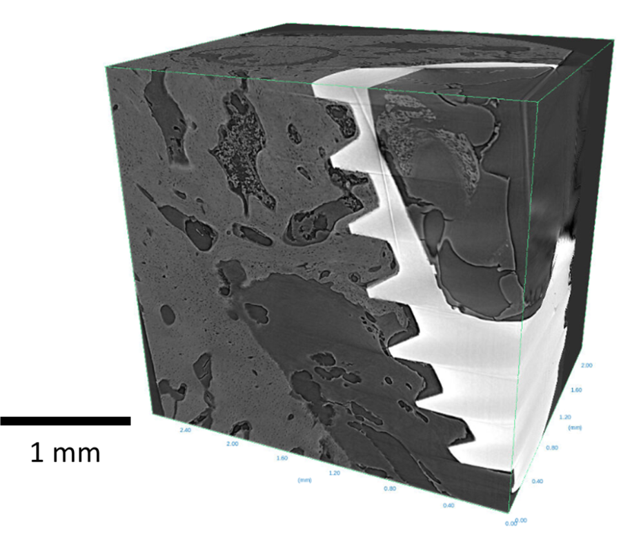
The Micro-Computed Tomography (MCT) beamline at the Australian Synchrotron was produced high resolution 3D images of osteocyte lacunae surrounding dental implants
Griffith University researchers investigated the biological process involved in healing after dental implant placements using imaging data from the Australian Synchrotron.
Dental implant placements often have lengthy healing periods and risks of other complications. The success of dental implant healing relies on bone tissue connecting with the surface of the implant in a process is known as osseointegration.
Osseointegration is dependent on tiny living cells that maintain the bone matrix, osteocyte lacunae. The arrangement of these cells allows the bone to adapt and remodel to dental implants.
The team comprising Dr Yuqing Mu, and Prof Dr Yin Xiao used the Micro-Computed Tomography beamline to generate high-resolution 3D images that revealed the structure of osteocyte lacunae around implants in animal bone tissue, during the osseointegration process.
"The MCT beamline can produce high resolution, three-dimensional images in micron size to visualise small things like osteocyte lacunae. It allowed researchers to see the healing between bone and the implant" explained Dr. Benedicta Arhatari, MCT beamline scientist.

By understanding the role of osteocyte lacunae in the healing process, scientists can improve design of implant surfaces and materials. This will improve the integration of dental implants, leading to better outcomes for patients.
"Researchers can take MCT images of several different implant material or surface roughness and see how the bone heals to decide which implant material and surface is best for bone healing" added Dr. Arhatari.
Implant samples in animals were exposed to synchrotron X-rays and rotated to collect projection data from multiple angles. The data from the MCT beamline was then reconstructed into 3D images using advanced software.
The MCT beamline is also able to produce phase contrast imaging. Phase contrast captures subtle variations in X-ray bending as they pass through a sample. This is very useful to visualise internal structures with low density like osteocyte lacunae.
In labs, it may take 12 to 13 hours to get these types of 3D images but, "the MCT beamline is able to produce these high-resolution images in just 10 minutes due to the brightness of the synchrotron beam," explained Dr Arhatari.
Related studies have been published in the journal Clinical Implant Dentistry and Related Research (2016) and Biomaterials (2019).
Thanks to Macy Huang, a student at ANU, who was assisted by Dr. Benedicta Arhatari in preparing this content.
Sample courtesy from Griffith's University, Dr Yuqing Mu and Professor Dr Yin Xiao.


