A bioengineering professor at The University of Texas at Arlington is developing a technique to diagnose tiny breast tumors that could reduce the anxiety, uncertainty and high costs often faced by patients.
Biopsy results show that about 80% of very small tumors are benign; 15% are low-grade, non-life-threatening cancers; and 5% are aggressive and invasive tumors that need immediate attention.
The National Institutes of Health recently awarded Baohong Yuan a three-year grant worth more than $440,000 to develop a method to use high-resolution imaging with super-sensitive temperature probes to determine if these tiny tumors are active and potentially harmful and, if so, to what degree.
"The majority of very small tumors may be nothing to worry about, but to know for sure costs a lot of money, takes a lot of time and leads to a lot of anxiety in patients," he said. "We hope that our method will allow doctors to diagnose and treat very small tumors more effectively without the need for a biopsy in every case, saving time and money and minimizing worry."
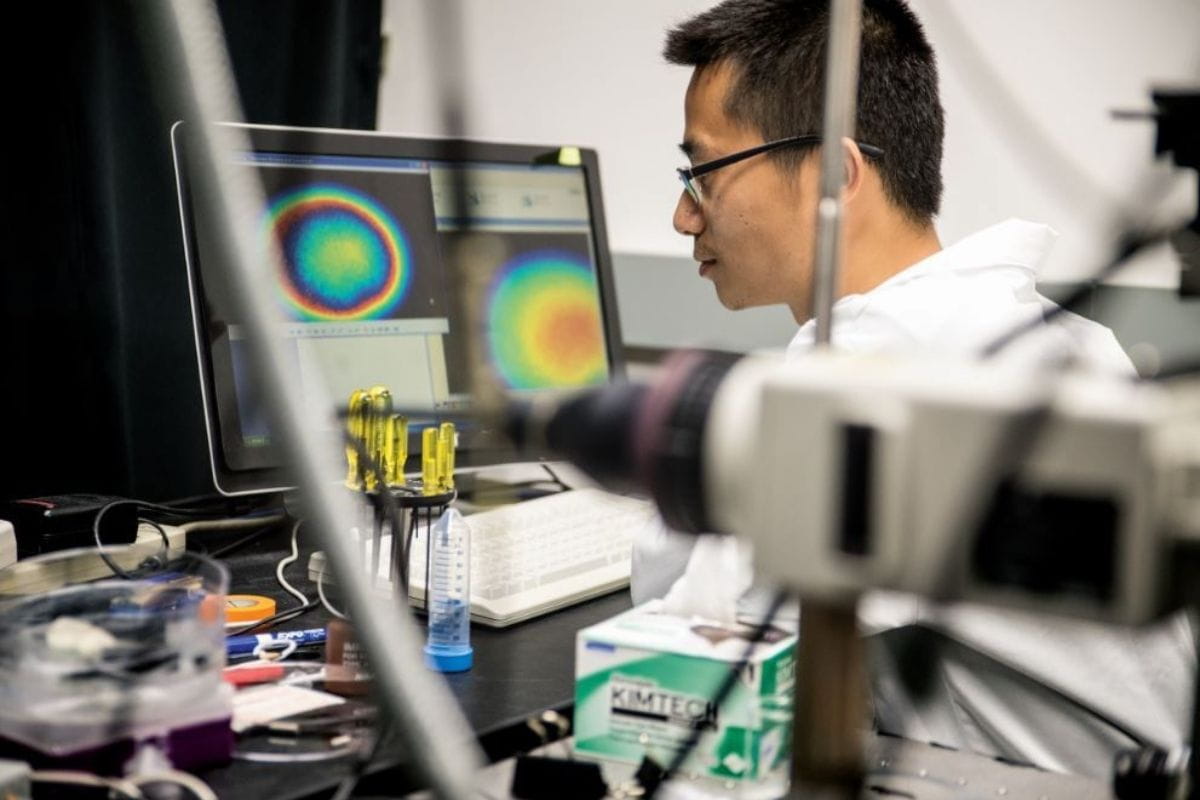
In active tumors, cells grow quickly and consume energy, which generates heat. Yuan said he hopes to use biocompatible nanoparticles with ultrasound to detect the temperature difference between the tumors and the surrounding tissue. The nanoparticles glow weakly at normal body temperature but increase in intensity at higher temperatures.
Once the nanoparticles reach the tumors via the bloodstream, they become activated by the tumor and ultrasound. Yuan says the increased intensity is both easily detected and provides tissue thermal information.
Using a process he developed for high-resolution imaging for deep tissue, Yuan can analyze the glow of the nanoparticles in the tumor to determine if it is active or if further observation or a biopsy are warranted.
The nanoparticles naturally pass out of the body, so the procedure will be safe for the patient.
When exposed to ultrasound waves, the particles become temporarily fluorescent and can be detected by a non-invasive probe system that he and other researchers designed. The technique enables researchers to see, measure, analyze and manipulate tissue in new ways, and resulting the information is useful in deciding treatment.
Yuan's work on deep-tissue imaging has had a significant impact on cancer diagnosis and treatment, says Bioengineering Department Chair Michael Cho.
"When successfully completed, his work can lead to overcoming multiple challenges of detecting small tumors and distinguishing active vs. inactive breast cancers," Cho said. "Solving the problem of overdiagnosis and overtreatment by new imaging technology can significantly minimize the adverse impact of cancer treatments."
- Written by Jeremy Agor, College of Engineering


