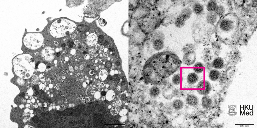
(Left) Low magnification electron micrograph of a monkey kidney cell (Vero E6) after infection with the SARS-CoV-2 Omicron variant showing cell damage with swollen vesicles containing small black viral particles.
(Right) High magnification electron micrograph of an infected Vero E6 cell showing aggregates of viral particles with corona shaped spikes on their surface (red box).
Photo credit: Professor John Nicholls, Clinical Professor of Department of Pathology; and Professor Malik Peiris, Tam Wah-Ching Professor in Medical Science and Chair Professor of Virology, School of Public Health, HKUMed; and Electron Microscope Unit, HKU.
/Public Release. This material from the originating organization/author(s) might be of the point-in-time nature, and edited for clarity, style and length. Mirage.News does not take institutional positions or sides, and all views, positions, and conclusions expressed herein are solely those of the author(s).View in full here.


