Most of us think little of why we feel pleasantly full after eating a big holiday meal, why we start to cough after accidentally inhaling campfire smoke, or why we are hit with sudden nausea after ingesting something toxic. However, such sensations are crucial for survival: they tell us what our bodies need at any given moment so that we can quickly adjust our behavior.
- By CATHERINE CARUSO
Yet historically, very little research has been devoted to understanding these basic bodily sensations-also known as internal senses-that are generated when the brain receives and interprets input from internal organs.
Now, a team led by researchers at Harvard Medical School has made new strides in understanding the basic biology of internal organ sensing, which involves a complicated cascade of communication between cells inside the body.
In a study conducted in mice and published Aug. 31 in Nature, the team used high-resolution imaging to reveal spatial maps of how neurons in the brain stem respond to feedback from internal organs.
They found that feedback from different organs activates discrete clusters of neurons, regardless of whether this information is mechanical or chemical in nature - and these groups of neurons representing different organs are topographically organized in the brain stem. Moreover, they discovered that inhibition within the brain plays a key role in helping neurons selectively respond to organs.
"Our study reveals the fundamental principles of how different internal organs are represented in the brain stem," said lead author Chen Ran, research fellow in cell biology at HMS.
The research is only a first step in elucidating how internal organs communicate with the brain. However, if the findings are confirmed in other species, including humans, they could help scientists develop better therapeutic strategies for diseases such as eating disorders, overactive bladder, diabetes, pulmonary disorders, and hypertension that arise when internal sensing goes awry.
"I think understanding how sensory inputs are encoded by the brain is one of the great mysteries of how the brain works," said senior author Stephen Liberles, professor of cell biology in the Blavatnik Institute at HMS and an investigator at Howard Hughes Medical Institute. "It gives inroads into understanding how the brain functions to generate perceptions and evoke behaviors."
Understudied and poorly understood
For almost a century, scientists have been studying how the brain processes external information to form the basic senses of sight, smell, hearing, taste, and touch that we use to navigate the world. Over time, they have compiled their findings to show how the various sensory areas in the brain are organized to represent different stimuli.
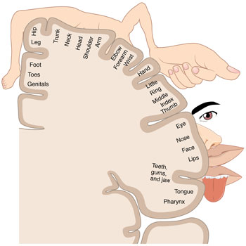
In the mid-1900s, for example, research on touch led scientists to develop the cortical homunculus for the somatosensory system-an illustration that depicts cartoonish body parts draped over the surface of the brain, each part positioned to align with the location where it is processed, and drawn to scale based on sensitivity. In 1981, Harvard professors David Hubel and Torsten Wiesel won a Nobel Prize for their research on vision, in which they methodically mapped the visual cortex of the brain by recording the electrical activity of individual neurons responding to visual stimuli. In 2004, another pair of scientists won a Nobel Prize for their studies of the olfactory system, in which they identified hundreds of olfactory receptors and revealed precisely how odor inputs are arranged in the nose and brain.
However, until now, the process by which the brain senses and organizes feedback from internal organs to regulate basic physiological functions such as hunger, satiation, thirst, nausea, pain, breathing, heart rate, and blood pressure has remained mysterious.
"How the brain receives inputs from within the body and how it processes those inputs have been vastly understudied and poorly understood," Liberles said.
This is perhaps because internal sensing is more complicated than external sensing, Ran added. External senses, he explained, tend to receive information in a single format. Vision, for example, is based entirely on the detection of light.
By contrast, internal organs convey information through mechanical forces, hormones, nutrients, toxins, temperature, and more-each of which can act on multiple organs and translate into multiple physiological responses. Mechanical stretch, for example, signals the need to urinate when it occurs in the bladder, but translates into satiation when it happens in the stomach and triggers a reflex to stop inhalation in the lungs.
A constellation of neurons
In their new study, Liberles, Ran, and colleagues focused on a brain stem region called the nucleus of the solitary tract, or NTS.
The NTS is known to receive sensory information from internal organs via the vagus nerve. It relays this information to higher-order brain regions that regulate physiological responses and generate behaviors. In this way, the NTS serves as an internal sensory gateway for the brain.
The researchers used a powerful technique called two-photon calcium imaging that measures calcium levels in individual neurons in the brain as a proxy for neuronal activity.
The team applied this technique to mice exposed to different types of internal organ stimuli and used a microscope to simultaneously record the responses of thousands of neurons in the NTS over time. The resulting videos show neurons lighting up throughout the NTS, much like stars winking on and off in the night sky


