Cornell researchers and collaborators have developed a neural implant so small that it can rest on a grain of salt, yet it can wirelessly transmit brain activity data in a living animal for more than a year.
The breakthrough, detailed Nov. 3 in Nature Electronics, demonstrates that microelectronic systems can function at an unprecedentedly small scale, opening new possibilities for neural monitoring, bio-integrated sensing and other applications.
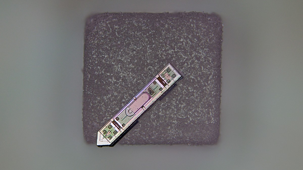
A neural implant developed at Cornell rests on a grain of salt. About 300 microns long and 70 microns wide, it's the smallest neural implant capable of wirelessly transmitting brain activity data.
Development of the device, called a microscale optoelectronic tetherless electrode, or MOTE, was co-led by Alyosha Molnar, the Ilda and Charles Lee Professor in the School of Electrical and Computer Engineering, and Sunwoo Lee, an assistant professor at Nanyang Technological University who first began working on the technology as a postdoctoral associate in Molnar's lab.
Powered by red and infrared laser beams that pass harmlessly through brain tissue, the MOTE transmits data back using tiny pulses of infrared light, which encode the brain's electrical signals. A semiconductor diode made of aluminum gallium arsenide captures light energy to power the circuit and emits light to communicate the data. Supporting this is a low-noise amplifier and optical encoder built using the same semiconductor technology in everyday microchips.
The MOTE is about 300 microns long and 70 microns wide.
"As far as we know, this is the smallest neural implant that will measure electrical activity in the brain and then report it out wirelessly," Molnar said. "By using pulse position modulation for the code - the same code used in optical communications for satellites, for example - we can use very, very little power to communicate and still successfully get the data back out optically."
The researchers tested the MOTE first in cell cultures and then implanted it into mice's barrel cortex, the brain region that processes sensory information from whiskers. Over the course of a year, the implant successfully recorded spikes of electrical activity from neurons as well as broader patterns of synaptic activity - all while the mice remained healthy and active.
"One of the motivations for doing this is that traditional electrodes and optical fibers can irritate the brain," Molnar said. "The tissue moves around the implant and can trigger an immune response. Our goal was to make the device small enough to minimize that disruption while still capturing brain activity faster than imaging systems, and without the need to genetically modify the neurons for imaging."
Molnar said the MOTE's material composition could make it possible to collect electrical recordings from the brain during MRI scans, which is largely not feasible with current implants. The technology could also be adapted for use in other tissues, such as the spinal cord, and even paired with future innovations like opto-electronics embedded in artificial skull plates.
Molnar first conceived of the MOTE in 2001, but the research didn't gain momentum until he began discussing the idea about 10 years ago with members of Cornell Neurotech, a joint initiative between the College of Arts and Sciences and Cornell Engineering. Co-authors of the paper include Chris Xu, director of the School of Applied and Engineering Physics; Paul McEuen, the John A. Newman Professor Emeritus in the Department of Physics (A&S); Jesse Goldberg, the Dr. David Merksamer and Dorothy Joslovitz Merksamer Professor in Biological Sciences in the Department of Neurobiology and Behavior (A&S); and Jan Lammerding, professor in the Meinig School of Biomedical Engineering.
The research was supported in part by the National Institutes of Health. Fabrication work was performed in part at the Cornell NanoScale Facility, which is supported by the National Science Foundation.
Syl Kacapyr is associate director of marketing and communications for Cornell Engineering.


