A 3D-printed titanium bone implant is giving a senior rescue dog a new 'leash' on life, thanks to a team of vets from The University of Queensland and engineers from RMIT University.
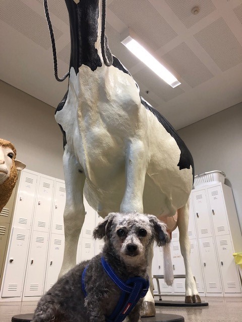
Seymour has gone from a sick rescue dog who was 'on his last legs' to a happy pooch who enjoys sunbaking on the deck.
Seymour was presented to Dr Jayne McGhie at UQ Vets Small Animal Hospital by his rescue adopter Sonya, looking to investigate options for a leg that may have been broken for many years.
"When Seymour first arrived in my care he had many problems - including severe skin disease, serious dental problems, dry eye, bilateral patella luxations and evidence of osteoarthritis in both elbows and in his right hind leg which required surgical intervention," she said.
 In the absence of standard solution Dr McGhie engaged Professor Milan Brandt, Technical Director of the Advanced Manufacturing Precinct at RMIT University in Melbourne, who, along with postdoctoral researcher Dr Darpan Shidid, donated his time and expertise to help.
Drawing on their current research on new generation implants for human bone disease, they designed and manufactured a custom-made, lattice-based titanium implant for Seymour using 3D metal printing technology at the Precinct.
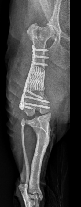
"After examining Seymour's CT scans, we designed a robust lattice structure that would support his weight and attached it to a custom-designed plate fitted exactly to his misshaped bone," Professor Brandt said.
"The lattice fills the bone defect to restore the femur to its normal length and alignment, while allowing growth of new bone as the femur heals - eventually the implant becomes a part of the healed bone."
Once Seymour had recovered from his skin and dental diseases, Dr McGhie and her team performed the advanced surgical procedure to install the implant.
"To encourage bone growth into the lattice, a bone graft was harvested from Seymour's shoulder," she said.
"This was mixed with canine demineralised bone and Seymour's own platelet rich plasma and then pressed into the lattice of the bone plate.
"The lattice and the plate were then placed into the bone defect of Seymour's left femur and secured in place."
 After his successful surgery, Seymour was sent home to recuperate with Sonya and her family.
"Seymour is such a remarkable little dog and despite all the challenges he's faced, he's never stopped being loving and happy," Sonya said.
"He's since had routine check-ups and - thanks to RMIT University and the specialist surgeon team at UQ VETS - is clinically doing very well".
"We're so happy with his recovery - he can do his excited twirls for food without a limp, and we can take him on walks with our other dog, Cash."
Image above left: Seymour's e-ray
Image above right: Seymour on the day of his surgery


