How to bring a diagnostic process that has endured for 100 years into the digital age? Two researchers from ETH Zurich and the University of Zurich are developing a robotic platform that enables a more accurate diagnosis of cancer cells by rapidly quantifying tissue samples in their entirety.
It all started with an innocuous question at the start of Francesca Catto's doctoral thesis: wouldn't it be nice if tissue samples could be coloured and digitally displayed as a 3D image? For over 100 years, histology, a branch of pathology that deals with tissue changes, has been using an analogue method that involves cutting tissue samples into micrometre-thin slices (about seven times thinner than a human hair) and examining them for pathological mutations under the microscope. This technique results in one in six people being misdiagnosed and cancer cells going undetected.
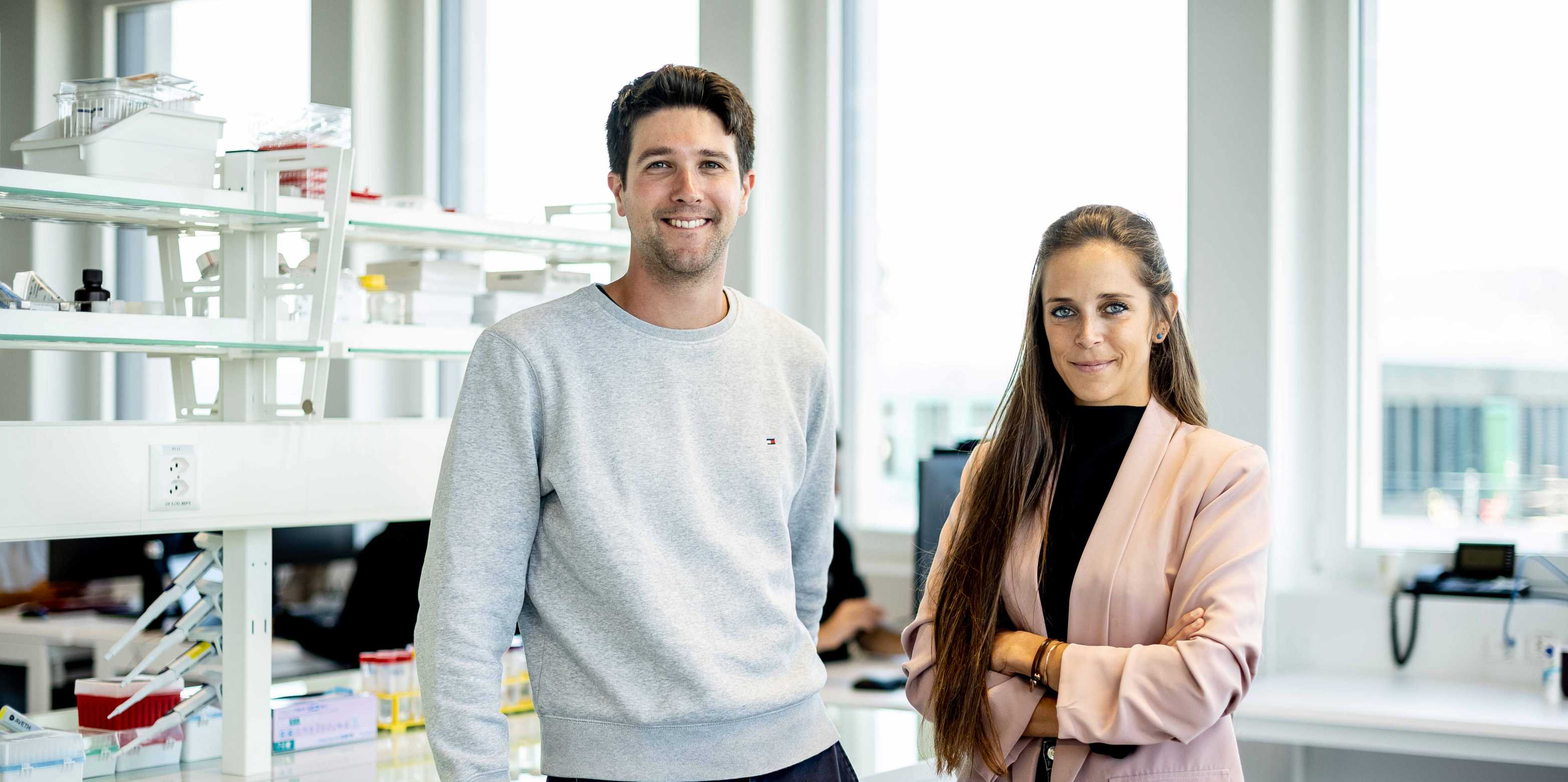
Catto, who did her thesis in neuroscience at the University of Zurich under the supervision of Professor Adriano Aguzzi, describes the early days as difficult: "We started out trying different approaches, but nothing worked. It was a nightmare. Fortunately, we entered into a collaboration with the groups of Professor Mirko Meboldt and Professor Alexander Mathys that resulted in the emergence of a successful approach. That's how Robert Axelrod, who has a doctorate in the field of processing technologies from ETH Zurich, joined the project."
Innovation through a groundbreaking interdisciplinary solution
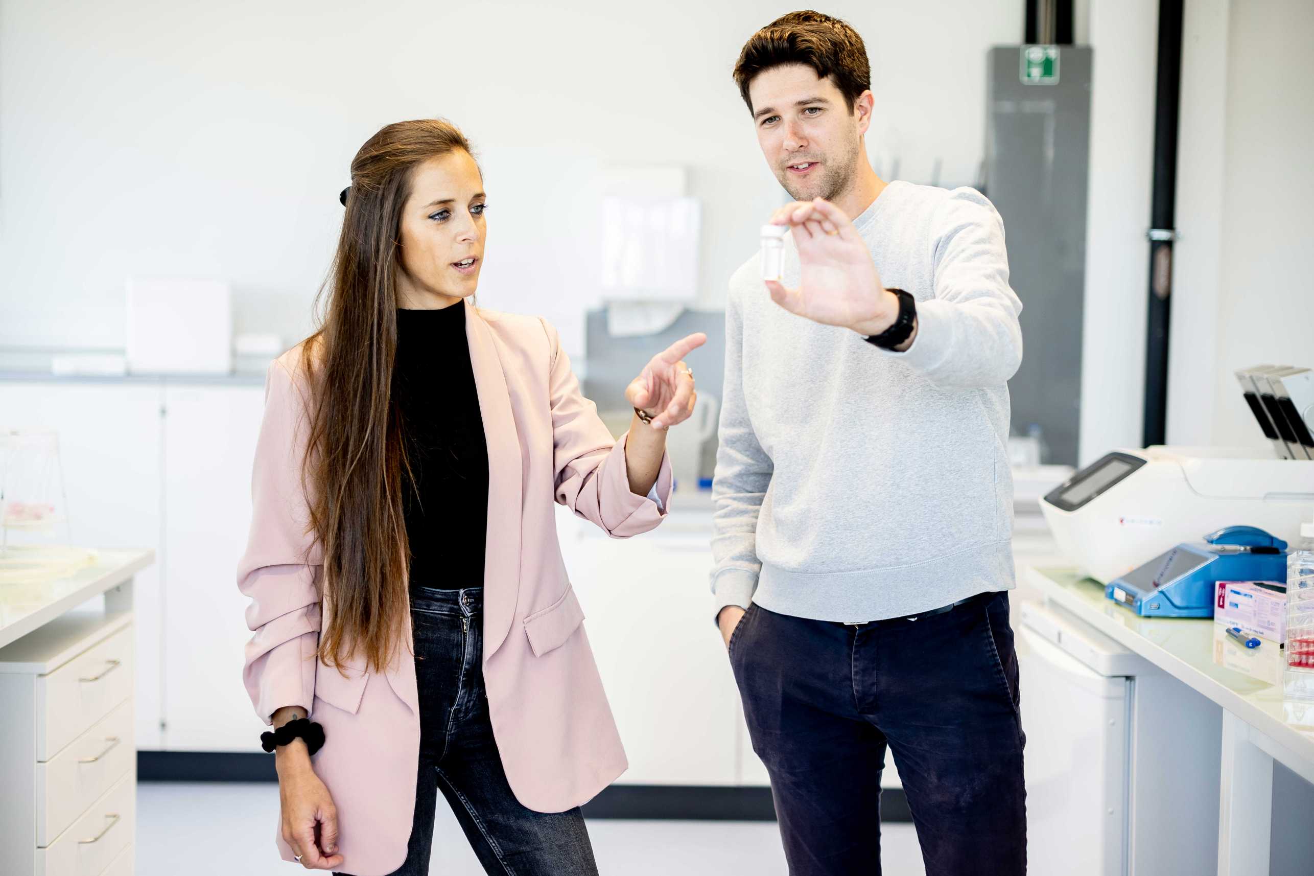
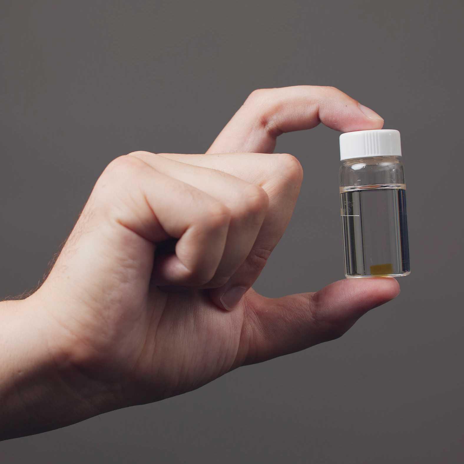
Using an approach that combines technologies from biomedicine and mechanical engineering, the two researchers developed a robotic platform that diagnoses cancer more accurately and provides three-dimensional information about the cells' spatial arrangement. This process comprises four steps. First, the researchers make the tissue sample transparent in an automated way. Next, they rapidly mark or colour any conspicuous cells. The third step is to create an image of the 3D tissue where cancer cells are mapped; the technology for this already exists. The final step is an analysis with 3D imaging software and training algorithms.
This innovative solution no longer calls for tissue samples to be prepared and cut in a time-consuming manner; instead, the tissue - a lymph node, say - is preserved whole and examined in its entirety. The digital 3D visualisation with the marked cells can be accessed online at any time.
New experiences and successes
In the lab, the robot prototype works and moves the samples around. However, the platform is not yet fully ready for the market. Axelrod says that while they can offer initial services by making sent-in tissue transparent in an automated way and quickly producing a labelled 3D image, they still need to optimise the software.
The two researchers also face challenges of a different kind as part of their Pioneer Fellowship, which is intended to lead to the founding of a start-up: "As a scientist, you have a different mindset when it comes to things like time management. Researchers are often perfectionists, because they don't publish anything until it's absolutely perfect and tested." But this project is allowing Catto and Axelrod to take their first steps in the world of start-ups, where they have to try out more, get feedback and go through fast, iterative cycles. "This has a major impact on time management," Catto says.
Catto and Axelrod cite their fantastic team as one of their biggest successes so far. Finding people with the right technical and social skills to get the team dynamic the two researchers wanted was a new experience for them both. Carrying out the full procedure for the first time was also an important technical milestone.
Always keep the goal in mind
When asked if there's time for hobbies besides the countless hours spent in the office and the lab, both of them laugh. The question seems obvious. For the time being, it's still possible to maintain a healthy work-life balance, they say, and that's important to them. "Although I fear that might change once the next phase of the project begins," Catto says. In difficult moments, they are motivated by the idea of their start-up doing well and the market launch of their product: "Thinking about the positive impact our robotic platform will have on histology is very motivating when times are tough. That's what drives us, especially whenever things don't go as planned," Axelrod adds.
The goal of bringing a good product to market is important to both of them - and not just because they personally want to see their start-up succeed. They also want to give research labs and clinics a useful, functional and reliable product that will catapult cancer diagnosis into the digital world.
Personal development is close to the duo's heart as well. The world of start-ups is, after all, the perfect place to become a good leader and still retain your own personality, to grow as a person and have new experiences.
References
Porcheri C, Ohad G, Calero‐Nieto FJ, Thambyrajah R, Ruiz‐Herguido C, Wang X, Catto F et al. (2020). Notch ligand Dll4 impairs cell recruitment to aortic clusters and limits blood stem cell generation. The EMBO Journal 39, no. 8: p.e104270. DOI: external page10.15252/embj.2019104270
Arcari M, Axelrod R, Adamcik J, Handschin S, Sánchez-Ferrer A, Mezzenga R, Nyström G. (2020). Structure-property relationships of cellulose nanofibril hydro- and aerogels and their building blocks. Nanoscale, 12(21), 11638-11646. DOI: external page10.1039/D0NR01362E


