
A new surface engineering approach that enhances mitochondrial stability and cellular uptake promises to advance organelle-based therapeutics.
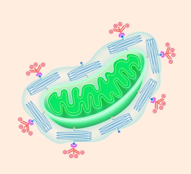
Inside almost every cell in our body are tiny structures called organelles that keep us alive and functioning. Among these are bean-shaped mitochondria, often referred to as the cell's power plants because they generate the energy our bodies need to survive. When mitochondria stop working properly, the consequences can be severe, contributing to diseases that affect the brain, heart, and other vital organs.
Many current approaches to treating mitochondrial disorders focus on supplying the key missing component, like a protein or antioxidant. But what if we could deliver mitochondria themselves directly into the target cells to modulate their energy metabolism?
This strategy, known as mitochondrial transplantation, has attracted growing interest, but it continues to face major technical challenges. Conventional mitochondrial preparations are fragile, they rapidly lose their function during handling and storage, and are also not easily taken up by cells.
To overcome these challenges, scientists at Hokkaido University have now developed a more efficient method for delivering mitochondria into target cells in a way that preserves their energy-producing function. The team described the new technology in a paper recently published in Advanced Materials Interfaces.
The researchers used a surface engineering approach that stabilizes mitochondria and enhances their ability to interact with cells. For this, isolated mitochondria are first coated with a protective layer made of polyethylene glycol (PEG), a material widely used in pharmaceuticals. This layer acts like a shield, helping to preserve mitochondrial structure while also serving as a platform for attaching functional molecules.
The team then added cell-penetrating peptides (CPP) to the outer end of this PEG layer. By positioning these peptides away from the mitochondrial membrane, this approach helped to enhance mitochondrial uptake and also preserved the integrity of the mitochondria's outer membrane under experimental conditions.
In laboratory experiments, the CPP-PEG-engineered mitochondrial preparations were seen to be taken up more efficiently by cells than unmodified mitochondria. And, the recipient cells for the modified mitochondria subsequently showed increased mitochondrial respiratory activity.
Researchers have named their new approach "e-MITO" for enhanced artificially designed mitochondria.
Professor Yuma Yamada, who led the study, says, "The modular nature of this approach could be a real advantage that could enable us to design mitochondria targeted to treat a specific disease or tissue. This work represents an early step toward a new field in which organelles themselves serve as therapeutic tools, much like genes, proteins, and cells. Organelle-derived materials could offer new opportunities to control cellular functions in the future."
Original article:
Shiraishi et al., CPP-PEG-Guided Surface Engineering of Mitochondria Enables Efficient Cellular Uptake and Respiratory Modulation. Advanced Materials Interfaces. July 2026.
DOI: 10.1002/admi.70583
Funding:
This work was supported in part by the Japan Science and Technology Agency (JST) through the Fusion Oriented Research for Disruptive Science and Technology (FOREST) Program (JPMJFR203X) and by the Japan Agency for Medical Research and Development (AMED; JP25ym012684 and JP223fa627005).


