
Epithelial cells are the tiny shields that line and protect our body. In a developing embryo, epithelial cells grow, divide, and move into positions to form the outer layers of our skin and the surfaces of our organs and blood vessels. When we scrape our skin, suffer an internal tear, or undergo surgery, epithelial cells will migrate to the site of injury to heal a wound. And when epithelial cells go haywire, they can turn malignant and spread through the body as cancer.
MIT engineers have now discovered that as they migrate, epithelial cells can synchronize and collectively pulse. In a study appearing today in the journal Newton , the researchers report observing groups of epithelial cells repeatedly moving in, then out, like a circle of dancers coming together and pulling apart.
The team measured this collective rhythmic pulsing in different types of epithelial cells, including healthy cells, cells from benign tumors, and cancerous cells.
Surprisingly, they discovered that malignant epithelial cells were more persistent in their synchronization, pulsing together for twice as long as healthier cells. It's unclear why the cells sync up in this way. But the researchers suspect that this cellular dance can serve as a clinical signal.
"More aggressive cancer cells tend to have a steadier and more persistent rhythm as compared to healthy ones," says study author Ming Guo, professor of mechanical engineering at MIT. "We think this coordination could serve as an early warning sign of how likely a tumor is to spread. The same coordinated waves may help shape embryos during development and close wounds upon injury."
The study includes first author and former MIT graduate student Wenhui Tang SM '20, PhD '24; Mehrana Nejad and L. Mahadevan of Harvard University; and Adrian Pegoraro of the Metrology Research Centre of the National Research Council Canada.
Cells got rhythm
When studying how epithelial cells organize and develop into whole organs and tissues, scientists have focused mainly on how the cells coordinate in space. Where cells move, where they are in relation to the growing tissue, and where they end up, are questions of spatial coordination that scientists including Guo have looked to investigate. How the movement of cells relate over time is less well-understood.
Guo's group at MIT studies cell interactions to identify patterns that relate to healthy versus diseased states. As part of this work, the team takes microscopic snapshots of cells that they grow in the lab, to identify interesting behaviors among cells. Recently, Tang, then a member of Guo's lab, was looking at a series of movies of epithelial cells when she started to see a rhythm, or pattern over time.
"I was studying collective cell migration, and I observed cells were swelling, then squeezing together, then swelling, again and again, forming local patterns," Tang recalls. "That's when I realized there might be something more interesting happening with these cells over time."
Taking a pulse
In their new study, the researchers focused on the timing of cellular movements. They started by studying healthy, live epithelial cells that they cultured in the lab. They stained the cells with fluorescent dye to illuminate each cell's nucleus. This way, they could easily identify one cell from another. They kept the cells in dishes with nutrients to help them naturally grow, divide, and move about.
"We're looking at their natural migration process, related to how they would migrate during different processes in the body, such as when forming skin and organs, and healing wounds," Guo explains.
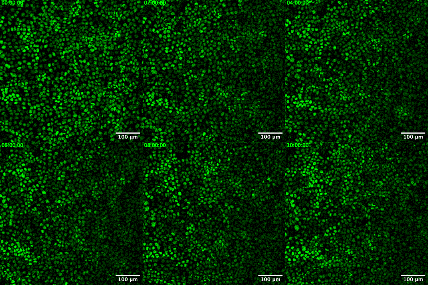
Using a confocal microscope, the team took snapshots of the cells every few minutes, for up to 30 hours. When they strung the images together as a sort of movie, a distinct pattern emerged.
Credit: Courtesy of the researchers
"If you just stare at any one location, you can see those dots are coming together, and then going further away, then coming together again, and going further away, like waves," Tang says.
They observed that a single pulse occurred over about an hour. This pulsing persisted in healthy cells, as a slow and steady rhythm over the 30-hour period.
Curious as to whether other types of epithelial cells would sync up in similar fashion, the team tried the same experiment with several different lines of human breast cancer epithelial cells. They studied the movement of cells from benign tumors and cells of increasing malignancy. They observed similar pockets of synchronized pulsing in every cell type, especially in the most cancerous cells.
"We found the really dangerous cancer cells team up over time, and do this persistent oscillation, twice as long as healthy cells," Guo says. "This is unexpected. We see they really team up, synchronize, and oscillate together, which potentially facilitates their invasion."
The researchers also observed a correlation between cell synchronization, and cell density: In each dish of cells, regardless of type, the cells continued to grow, divide, and pulse. As their numbers grew, more cells pulsed together, and their synchronization increased, up to a point. Once the cells reached a certain density, their pulsing began to die down.
"There's a peak of synchrony before it decreases as cell density continues to increase," Tang says.
This connection is especially interesting in the context of certain conditions such as asthma. Epithelial cells line the inside of many organs and tissues, including the airways. In healthy people, these cells pack together and "jam" up to form a solid, stable lining that protects the airways. In asthmatic airways, however, epithelial cells are less able to jam together. This results in airways that are loose and fragile, easily irritated, and difficult to heal.
Guo and Tang suspect that, as there appears to be a connection between cell density and cell synchronization, there may be a way to target asthma treatments, by watching how potential drugs affect asthma cell synchronization. A similar approach could be taken for the screening of cancer drugs.
"More malignant cells would be better synchronized. After treating them with a drug, if their synchronization is disrupted, then it might be an efficient drug where we can consider the next step," Guo envisions.
This research was supported, in part, by the National Institutes of Health.


