An international collaboration study reveals how evolution and locomotion patterns, such as bipedalism, shaped bone structures through proteins present in the bone matrix. The findings of the study, led by researchers from the University of Turku, the UAB and the URL, are important for understanding bone fragility and regeneration, and could help guide the development of biomaterials inspired by natural skeletal adaptation.
A new international collaboration sheds light on the evolutionary mechanisms behind bone mechanoadaptation and highlights several protein families potentially involved in bone remodeling. The research was recently published in Communications Biology, a journal from the Nature group, and was led by researchers Pere Puigbò, from the Universitat Autònoma de Barcelona (UAB), and Miho Nakamura, from University of Turku (UTU, Finland) and La Salle Campus Barcelona, founding member of the Universitat Ramon Llull (URL).
Vertebrate bones constantly sense and respond to mechanical forces, a phenomenon known as mechanoadaptation. This new study shows that this ability has been shaped not only by biomechanics but also by millions of years of evolutionary pressure related to locomotion. Species with different locomotor patterns display distinct evolutionary signatures in the genes and proteins associated with load sensing and skeletal remodeling.
"These results indicate that the evolution of locomotion has played a major role in shaping the molecular machinery of bone mechanoadaptation," says Prof. Pere Puigbò. Two of these decisive moments were the transition of vertebrates from water to land, which increased the pressure on the limbs, and the emergence of bipedalism in humans, which redistributed stress between arms and legs.
The research identifies several non-collagenous proteins of the bone matrix that may act as key regulators of mechanotransduction — many of which have received limited attention in previous skeletal studies. These findings provide new insights into how bone cells detect and respond to forces, and how these mechanisms evolved across vertebrates.
"From a cell biology perspective, our work points to important but underappreciated proteins that could be central to bone remodeling," adds Prof. Miho Nakamura.
The findings are important for understanding bone regeneration, fragility, and osteoporosis, and could help guide the development of biomaterials inspired by natural skeletal adaptation. The study highlights several non-collagenous proteins, including Fetuin-A, which controls mineral deposition and prevents abnormal calcification. Fetuin-A's conserved role is key for maintaining healthy bones and may affect osteoporosis risk by balancing bone formation and breakdown.
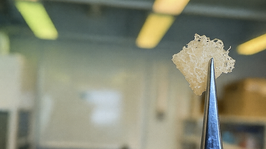
The study, conducted on bone cell cultures and using phylogenetic analyses, was funded by the Sigrid Jusélius Foundation, Finland, and the Japan Society for the Promotion of Science.
Building on these discoveries, the Phylobone research group, coordinated by both researchers and whose mission is to study bone regeneration in the light of evolution, is investigating how such proteins drive skeletal adaptation and bone remodeling.
Link to the original article: Shimochi S, Brunet C, Fontcuberta-Rigo M, Hrovat K, Puigbò P, Nakamura M. Bone mechano-response is driven by locomotion transitions during vertebrate evolution. Communications Biology (2025). https://doi.org/10.1038/s42003-025-09292-1


