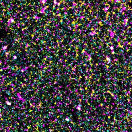
Figure 1: A microscopy image showing the distribution of epidermal growth factor receptor (EGFR; cyan) and PI(4,5)P2 (magenta) in a cell membrane. Modified from Ref. 1 and licensed under CC BY 4.0 © 2025 M. Abe et al.
Tiny blobs of fats, or lipids, in cell membranes regulate the activity of a cancer-linked protein, cell biologists at RIKEN have discovered1. This finding could pave the way for novel approaches for treating cancer.
Lying within the thin, flexible membranes that encase cells are tiny clusters of specialized lipids that help to steer critical growth signals. But just how these structures, known as nanodomains, coordinate these complex processes has long been a mystery.
The key role nanodomains play in regulating the activity of the epidermal growth factor receptor (EGFR)-a protein implicated in cell growth and division and also cancer-has now been uncovered by a team led by bioimaging specialist Yasushi Sako of the RIKEN Cellular Informatics Laboratory.
The RIKEN team captured the intricate choreography between lipid nanodomains and EGFR in living cells using super-resolution microscopy, which was capable of imaging single molecules.
The nanodomains-some of which contain a charged fat molecule called 'PI(4,5)P2'-activate EGFR by orchestrating key molecular interactions. After completing this task, the nanodomains dismantle themselves, thereby deactivating EGFR. This ensures precise and dynamic control of cellular signaling.
"The PI(4,5)P2 nanodomain supports EGFR activation, while activated EGFR dissolves the nanodomain structure," explains lipid biologist Mitsuhiro Abe, also of the RIKEN Cellular Informatics Laboratory.
This dynamic remodeling of the lipid environment reveals a previously underappreciated mechanism of signal regulation, where lipids in the membrane actively shape protein function to fine-tune cellular responses. This finding challenges the view of the cell membrane as a passive boundary. Instead, the membrane emerges as an active regulator, shaping how signaling proteins function to maintain cellular balance and control.
Nanodomains are far too tiny to be observed with conventional fluorescence microscopy. And so the team employed advanced single-molecule localization microscopy to track interactions between EGFR and PI(4,5)P2 lipids with unprecedented precision.
"This research was accomplished by combining the fields of lipid biology and cell biophysics," notes Abe. This interdisciplinary approach allowed the team to precisely observe how nanodomains cluster around EGFR before stimulation, enhancing its activation.
The team's discovery opens the door to developing new therapeutic strategies for cancer and hereditary syndromes, for which EGFR and related signaling proteins are often implicated. The lipid-mediated regulation uncovered by the team suggests that targeting not just EGFR, but also the lipid environment controlling it, could offer a novel approach to addressing tumors driven by aberrant signaling, potentially improving treatment outcomes.


