People in Northern California struggling with forms of peripheral artery disease (PAD) now have an alternative to open bypass surgery to treat their condition.
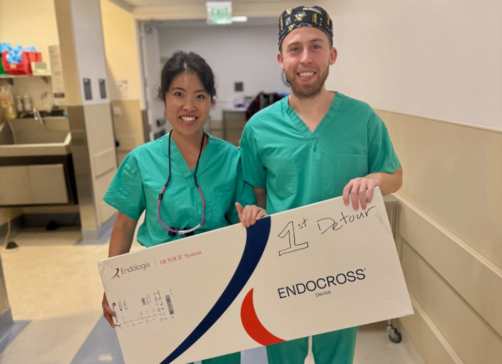
Vascular surgeons at UC Davis Medical Center recently completed their first percutaneous transmural arterial bypass therapy (PTAB) using the DETOUR System. UC Davis is the first hospital in the Sacramento region to perform this minimally invasive treatment.
"We are excited to offer our patients with complex forms of PAD this clinically proven and effective treatment," shared Melissa R. Keller, assistant professor of vascular surgery. Keller oversees the treatment of PAD and limb salvage for UC Davis Health. "This next-generation device allows us to provide an alternative treatment option with comparable outcomes to surgical bypass while avoiding potential bypass complications."
"This next-generation device allows us to provide an alternative treatment option with comparable outcomes to surgical bypass while avoiding potential bypass complications."-Melissa R. Keller

What is PAD?
PAD is a common condition in which narrowed arteries reduce blood flow to the arms or legs. This may cause leg pain when walking, weakness and poor healing.
The condition is usually a sign of a buildup of fatty deposits (plaque) in the arteries, a condition called atherosclerosis. This can cause arteries to narrow, blocking blood flow.
Complications from PAD caused by atherosclerosis can include:
- Blood clots
- Bloodstream infections
- Bone infections
- Critical limb ischemia (an injury or infection that causes tissue to die)
- Amputation
- Stroke and heart attack
The superficial femoral artery (SFA), which runs the length of the thigh, is one of the arteries most affected in patients with PAD.
For patients with blockages of the SFA, open surgical bypass is the current standard of care, but the procedure carries risks of complications and a prolonged recovery time.
About the procedure
PTAB is a minimally invasive approach that utilizes an endovascular technique using a patient's own blood vessels to create a bypass around the diseased or blocked area.
During the procedure, a stent graft is placed through a small hole in the patient's groin. Using a form of X-rays, the stent graft is guided from the femoral artery (the main artery in the leg) to the popliteal artery (a continuation of the superficial femoral artery) by way of the femoral vein. By using the femoral vein as a conduit, the diseased SFA is bypassed, allowing unobstructed blood flow around the blockage.
"This minimally invasive approach allows us to detour around even a long blockage in the artery without making a single cut on the affected leg."-Mimmie Kwong

"This minimally invasive approach allows us to detour around even a long blockage in the artery without making a single cut on the affected leg," explained Mimmie Kwong, assistant professor of vascular surgery, who performed the procedure.


