In a study published in Nature Methods on December 2, a research team led by Profs. XU Tao and JI Wei from the Institute of Biophysics of the Chinese Academy of Sciences has developed a three-dimensional interferometric localization microscope called repetitive optical selective exposure in 3D (ROSE-3D), achieving camera-based isotropic nanoscale resolution in 3D.
By designing a high-speed illumination-switching pathway based on an electro-optic deflector, the researchers enabled ROSE-3D to introduce interferometric light simultaneously along the X, Y, and Z axes, achieving an interference fringe switching time of less than 1 microsecond.
Through 3D interferometric localization, ROSE-3D overcomes dependence on single-molecule image shape. Within a depth of field of approximately one micrometer, the system improves lateral localization precision by 2-6 fold and axial precision by 3.5-8 fold. ROSE-3D single-layer imaging is capable of resolving 1-micrometer-thick intracellular structures with nanoscale resolution.
This study further extends ROSE-3D to multicolor and multilayer imaging, enabling dual-color visualization of the nuclear lamina proteins lamin A/C and lamin B1 in COS-7 cells. Quantitative analysis revealed that lamin A/C is positioned on the inner side of lamin B1, with an interlayer spacing of approximately 10 nm. This is the first time this spatial relationship has been revealed at the full-nucleus, 3D cellular scale.
In addition, leveraging its isotropic nanoscale resolving power, the researchers captured the in situ assembly of the mitochondrial fission protein DRP1 into multiple architectures on the mitochondrial outer membrane. This represents the first structural characterization of DRP1 assemblies within their native cellular environment.
These findings demonstrate that ROSE-3D, as a new 3D multicolor nanoscopic imaging technology, enables ultrahigh-resolution localization and structural analysis of subcellular organelles and macromolecular complexes, providing robust technical support for studies of in situ 3D cellular nanoarchitecture.
ROSE-3D holds significant promise for resolving diverse 3D nanoscale structures in biological samples and for uncovering the native assembly mechanisms of biomolecular complexes.
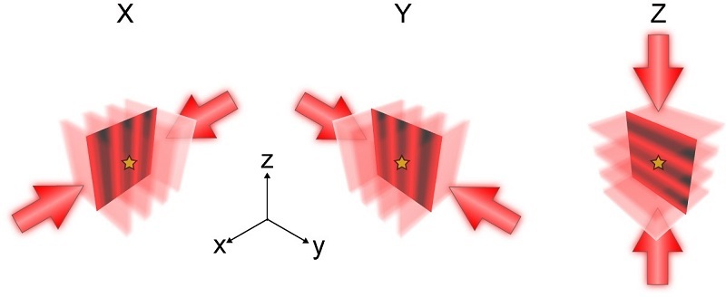
Figure 1. Principle of ROSE-3D (Image by XU Tao's group and JI Wei's group)
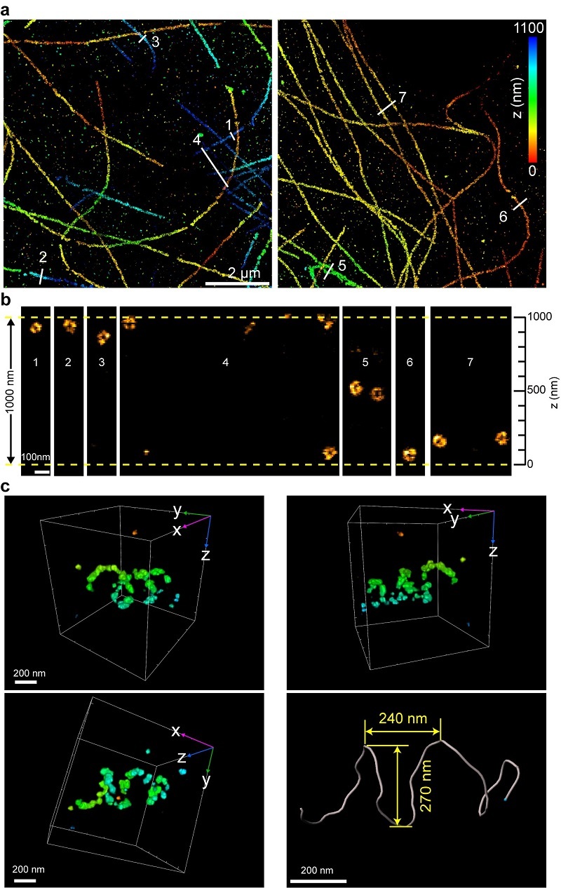
Figure 2. a-b. ROSE-3D resolved the hollow structure of microtubules; c. the helical structure of DRP1 complexes (Image by XU Tao's group and JI Wei's group)


