The knee joint is the largest and most complex weight-bearing joint in the human body that enables us to stand and carry out our daily tasks. How much do you know your knee? Human body is the most perfect masterpiece of nature, and the knee joint inside human body has a delicate ultrathin layer of osteochondral interface between SOFT cartilage and HARD subchondral bone. In this interface, the sophisticated structures and excellent force transfer properties allow the knee to resist fatigue damage over a lifetime of loading cycles. The interface's secret to successfully preventing such damage should lie in its well-designed micro-nano structures and graded compositions in multiple levels. Thus, revealing this ultrathin interface is key to understanding its superb mechanics and for the future design of soft-hard composite interface materials.
Against this backdrop, the team led by OUYANG Hongwei at the Zhejiang University School of Medicine conducted a high-resolution analysis of the osteochondral interface in human knee joints. It is the first time that researchers identified the osteochondral interface tissue from the perspectives of microstructure, component assembly and tissue mechanics. Moreover, they discovered the mechanism for the super-strong force transfer and fatigue-resistant adhesion of the ultrathin osteochondral interface. They identified an ultrathin ∼20−30 μm graded calcified region with two-layered micronano structures of osteochondral interface tissue in the human knee joint, which exhibited characteristic biomolecular compositions and complex nanocrystals assembly. Results from finite element simulations revealed that within this region, an exponential increase of modulus (3 orders of magnitude) was conducive to force transmission.
Their research findings were published in an article entitled "Identification of an Ultrathin Osteochondral Interface Tissue with Specific Nanostructure at the Human Knee Joint" in the journal Nano Letters.
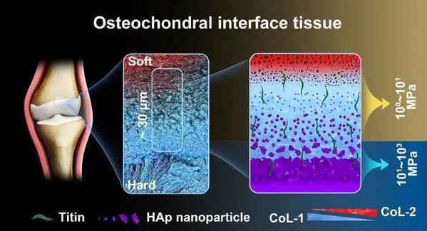
In their study, the researchers collected normal cartilage tissues and identified the osteochondral interface tissue via histological staining. Using scanning electron microscopy (SEM) and energy dispersive X-ray (EDX) line scan, they obtained an ultrathin graded calcified region, spanning over 20-30 μm. By analyzing the transition of the microstructure at the interface and the assembly pattern of HAp, they found the shift from the porous structure to the dense one. Meanwhile, HAp showed variations in morphology across the interface, denoting the maturity of the assembly. The spatially graded distribution of HAp was beneficial to reducing stress concentration and promoting force transmission.

Microstructure transition of the osteochondral interface
The schematic view of the osteochondral interface indicated two-staged modulus increments, particularly the 3 orders of magnitude increment in the 30 μm spatial range. The FEA results further demonstrated that this modulus transition facilitated mechanical conduction.
Biomechanics of the osteochondral interface tissues
The tissue modulus map is intimately bound up with the two-layered micronano structure variation at the interface. In addition to the structure, the gradient shift of compositions at the interface can also modulate its mechanical function by redistributing stress. Therefore, the researchers further examined the compositional assembly of the interface at multiple scales. Using XRD and Raman spectroscopy, they found that inorganic nanocrystals at the osteochondral interface were dominated by carbonated substituted HAp. With the extension of the interface, the carbonated substitution rate decreased, mineral crystallinity increased, the HAp composition gradually increased, and the calcium-phosphorus ratio increased from 1.2 to 1.6. These implied that HAp became gradually mature at the interface. With the help of HRTEM, SAED and ELLS, the heterogeneity of HAp crystal assembly was confirmed at the nanoscale. HAp with nanoscale heterogeneity turned out to be insensitive to cracking and thus could promote force conduction through energy dissipation.
Compositional analyses and nanoscale heterogeneity of HAp at the osteochondral interface
Besides, the researchers examined the precise protein expression profiles at the interface using LC-MS / MS and found that the interface tissue had a high expression of elastic-responsive protein-titin, which could absorb energy through reversible deformation and transmit stress, thereby helping maintain elasticity and mechanical conduction at the interface.
Quantitative proteome analysis of the difference in expression of
osteochondral interface compared to AC and SB
"A combination of characterization of microstructural, micromechanical, nanocompositional, and biomolecular features of the interface revealed the mechanism underlying toughening properties of the cartilage-to-bone interface tissue," said Prof. Ouyang. "The identified mechanism of the soft-to-hard interface allows effective force transfer in a certain direction, thus laying a foundation for future approaches seeking to design biocomposite interface materials."


