A new technique developed at the School identifies complications of rare heart problem, anomalous coronary artery
Researchers from the School of Biomedical Engineering & Imaging Sciences have used multimodal imaging with MRI and CT, including a new technique developed at the School, to identify complications of rare heart problem, anomalous coronary artery, in a man in his 20s. After undergoing corrective surgery, follow-up tests revealed the man is now clear of heart problems.
In the case report, published in JACC: Case Reports, the researchers identified a young patient in his 20s with no underlying health problems, who was having small episodes of heart attacks from the abnormal connection of the coronary artery.
Anomalous coronary artery is when arteries come out of the heart vessel in an abnormal position, affecting around 1 percent of the population.
Although they are present at birth, ACAs are often not diagnosed until late adolescence or adulthood, because of the lack of symptoms or because symptoms may not be recognized as being caused by ACA.
In this case study, the CT scan was performed as part of the patient's clinical care. He had an MRI scan as a result of the CT scan and additional imaging research was performed on the patient.
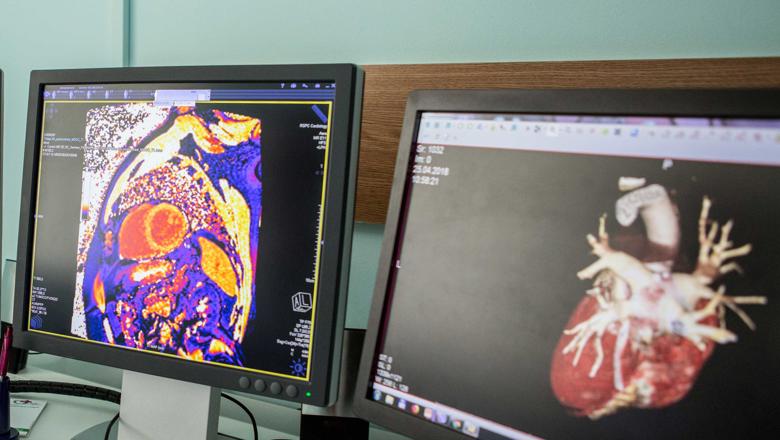
The researchers used MRI to detect the impact on the heart in a novel way, in particular with a new technique developed by Professor Amedeo Chiribiri, Professor in Cardiovascular Imaging and Consultant Cardiologist. The technique is called novel dark blood imaging for detecting scarring as with normal methods, small amounts of the scars cannot be identified.
With this new method implemented, the researchers demonstrated that using CT, which has an excellent image quality for high resolution imaging with the arteries, there was an abnormal origin of one of the coronary arteries. Using MRI, which is excellent for looking at the soft tissue of the heart, the researchers were also able to identify the impact of the problem on the heart and guide subsequent clinical decision making.
Professor Amedeo Chiribiri, Professor in Cardiovascular Imaging and Consultant Cardiologist, said clinicians have been using contrast in Cardiac MRI to assess for 'scarring' of the heart for over 20 years.
We have then realised that we were not very sensitive for the detection of small areas of scarring, which are still very relevant to guide patients' management. In collaboration with Professor Rene Botnar and Dr Robert Holtackers, from the Department of Biomedical Engineering at King's College London and Maastricht University in the Netherlands, we have designed a novel acquisition method which solves this shortcoming of standard methods.– Professor Amedeo Chiribiri, Professor in Cardiovascular Imaging and Consultant Cardiologist
"This new approach has the advantage of being immediately available on most MRI scanners worldwide without any additional software or hardware upgrades. Using this technique we have been able to detect and see greater amounts of scarring of the heart that we haven't been able to previously appreciate."
Lead researcher Dr Sohaib Nazir, NIHR clinical lecturer and cardiology registrar at King's College London and Guy's and St Thomas' Hospital said the team would not have picked up the disease and the complications of the anomalous coronary artery if they had not done the advanced imaging tests.
It is important to highlight to clinicians of the need for accurate diagnosis using the advanced imaging tools that we have at our disposal. If the patient had not done the CT scan at the initial stage, we would not have identified the issue in this young man. In addition, by using the novel MRI method we would not have appreciated the significance of the anomalous coronary artery. As a result, the findings from our imaging directly provided evidence of the need for the patient to undergo correct heart surgery. Finally, this is very much a team effort with collaboration with clinicians and specialists including in multimodal imaging including expertise from Dr Nabeel Sheikh and Dr Rebecca Preston from Guy's and St Thomas Hospital.– Dr Sohaib Nazir, NIHR clinical lecturer and cardiology registrar at King's College London and Guy's and St Thomas' Hospital


