Scientists have shown how the edges of biological tissues create geometric constraints that help cells position themselves in a magnet-like manner, giving rise to structure and order inside developing embryos
Summary
- During embryonic development, cells with biochemically distinct ends - called polarised cells - organise into distinct patterns based on their orientation.
- Using theoretical physics approaches and quantitative experiments, scientists have shown how the shape of tissue boundaries determines these patterns.
- They found that specific features in these patterns - called topological defects - determine where the proamniotic cavity, an important structure for embryonic development, forms.
- This research identifies fundamental principles that govern how order and functional structures emerge inside complex biological systems.
One of the most striking biological transitions in nature happens early in development, when an embryo transforms from a simple ball of cells into a highly ordered structure with distinct tissue layers that later develop into various organ systems. If one imagines the cells of an embryo as people, it is as if a disorderly crowd resolves spontaneously into neat rows and columns.
New research by EMBL researchers and their collaborators uses a combination of theoretical physics and experimental biology to show how this process arises from interactions between tissue geometry and the directional properties of cells. The results were published recently in companion publications in the journals Nature Physics and Nature Materials .
The studies resulted from a collaboration between Anna Erzberger's group at EMBL Heidelberg and Takafumi Ichikawa at Kyoto University, Japan, together with scientists at the Hubrecht Institute, Netherlands. Their goal was to understand how order emerges in complex biological systems.
When cells behave like tiny magnets
Pamela Guruciaga, a postdoc in the Erzberger Group, has long been interested in understanding how order develops in interaction with the environment. A physicist by training, she was struck by the similarities between the properties of certain biological systems and the magnetic systems she studied during her PhD.
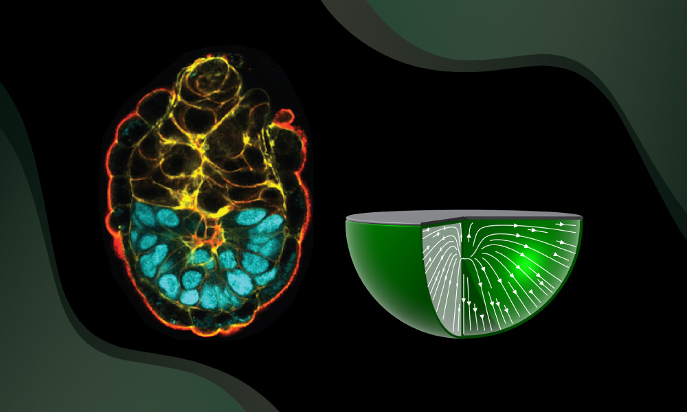
Certain cells are distinctly polarised - they have two ends that are very different from each other. In this, they resemble tiny magnets, which have north and south poles. In an early-stage mouse embryo, cells of the epiblast - which eventually give rise to all the major tissues - behave in this manner. These cells have an apical and a basal end, characterised by different compositions and concentrations of various biological molecules.
The team decided to investigate the fundamental principles governing the behaviour of systems where such polarised cells are present in bulk and face physical constraints at the tissue borders.
For example, imagine if people inside a crowded room were given certain rules as to which way they could face relative to the walls and floor and used this to orient themselves in relation to one another. Guruciaga and her colleagues wanted to figure out what these rules were for epiblast cells.
By focusing on how the cellular orientations interact with each other and their environment, the researchers built a minimal model that predicts how organisation changes when these interactions are altered.
To test their predictions, the team relied on experimental measurements led by Ichikawa, a researcher at Kyoto University, who began this collaboration as a postdoc in the lab of Takashi Hiiragi at EMBL (now a Group Leader at the Hubrecht Institute and a Visiting Professor at Kyoto University). While they were at EMBL, Ichikawa and colleagues had developed an ex vivo embryo culture system that allowed scientists to make precise three-dimensional measurements and see the effects of perturbations.
"It was a very synergetic and fun collaboration across time zones," said Guruciaga. "For me, as a physicist, I may know why something works, but it's still kind of magic to see that it's all true in messy biological systems. It was also super interesting coming from a pure physics perspective to come up with a common language to work with biologists."
On (topological) defects and development
The team observed experimentally that in the cup-shaped epiblast, different boundaries resulted in different orientations for epiblast cells. When the boundary was lined with the extracellular matrix - the three-dimensional network of proteins that surrounds some tissues - the cells oriented perpendicularly, with their basal ends towards the extracellular matrix. On the other hand, when the epiblast directly contacted a neighbouring tissue without such a matrix, the cells aligned parallel to the boundary.
The scientists found that the combination of these two orientations can result in an interesting phenomenon, the appearance of structures called 'topological defects'. "These are points in space where it is undefined in which direction an object should point," explained Guruciaga. "For example, if a set of arrows is arranged in a starburst pattern, the centre is a point where all directions are equivalent. These points are super relevant because they are very robust; you cannot easily destroy them."
Topological defects are known to control the collective properties of anisotropic systems - systems where all directions are not equivalent - in fundamental ways. This is similar to how charged particles can determine the behaviour of an electric field. They have also been shown to drive important functions in biological systems; for example, topological defects can trigger the formation of structures such as fruiting bodies in bacteria or tentacles in animals like Hydra.
However, previous studies focused on two-dimensional cell sheets, and the role of topological defects in the context of three-dimensional tissues made up of polarised cells had not been explored. The new theoretical framework developed by Guruciaga and her colleagues is one of the first to achieve this.
Importantly, for this three-dimensional system, Guruciaga discovered that the number and type of defects can be controlled by the geometry of the confining boundaries alone. This result shows that shape itself can determine the organisation of topological defects and associated structures. In embryos, this principle provides a robust mechanism for spatially controlling tissue organisation, one that is likely relevant in other biological systems as well.
In their experiments with the developing epiblast, the scientists indeed observed topological defects. Imaging embryos across developmental stages, they saw that early on, there were no defects, but as development progressed, a defect appeared at a particular stage. "This transition suggests that surface interactions become stronger," said Guruciaga. When the scientists measured the levels of some of the molecules that determine the strength of boundary interactions, they saw that these indeed increased as embryos grow and develop.
Predicting the time and place of lumen formation
Interestingly, the sites where these defects appeared corresponded to the positions where fluid-filled cavities, called lumina, later formed. "It was exciting to see that these topological features directly corresponded to where lumina emerged," said Guruciaga.
"The lumen eventually grows to become the pro-amniotic cavity, a crucial step in mammalian embryonic development," said Ichikawa. "We had long wondered what determines where it forms."
To directly test whether the boundary shape controls the number of defects as predicted by their theory, the scientists performed experiments where they altered the geometry of the epiblast. Indeed, consistent with the theory, perturbing embryo shape induced the formation of additional lumina at the predicted positions. This resulted in embryos that had two lumina, instead of just one, as in unperturbed embryos.
The scientists also saw that such boundary interactions and resulting cell orientations actively drive the maturation of the epiblast. "This interdisciplinary collaboration, which began at EMBL, gave us a physical explanation for how lumen formation is controlled in space and time - something we could not have arrived at from biology alone," said Ichikawa.
"What I find most exciting is that these results identify a very general physical principle," says Erzberger. "We show that geometry alone can determine orientation patterns in three dimensions, independent of the microscopic details of the system. That means shape itself can act as a robust control parameter - not just in embryos, but across a wide range of biological and physical systems."


