It sounds like science fiction - but lasers beating to the rhythm of a live heart is exactly what researchers at the University of St Andrews have developed to improve the understanding of heart failure and to help develop more effective treatments.
Leading an interdisciplinary team of scientists, Dr Marcel Schubert and Professor Malte Gather of the School of Physics and Astronomy and Dr Samantha Pitt of the School of Medicine at the University of St Andrews, embedded tiny lasers into individual heart cells, and by analysing the light these lasers produce they monitored the contractions of the heart muscle.
The research, published in Nature Photonics today (Doi: 10.1038/s41566-020-0631-z) comes in the year in which the laser marks 60 years since it's invention.
To check the function of the heart, doctors take a patient's pulse, measure blood pressure, or take an electrocardiogram (ECG) which provides information on the function and the rhythm of the heart as a whole, but provides little information about what happens in the different parts of the heart.
Echocardiograms and other sophisticated methods can provide more local information, but further advances, in particular for treatments which explore stem cells or transplanted tissue, will require to follow the contractions of the individual cells forming the heart muscle.
Achieving this, at least in animal models routinely used to study critical heart conditions commonly seen in human patients, promises improved understanding and thus more effective treatment.
Lasers are widely used in biomedical imaging, resolving ever finer details of life, including mapping details in heart cells. But because lasers are usually big and power-hungry, they sit outside the heart and can only send their light to the surface of the biological tissue, which severely limits how deep they can look.
In this latest work, tiny lasers were placed inside the heart where they acted as microscopic probes. With every beat of the heart, the colour of light that these lasers emit changed by a small but clearly detectable amount, thus precisely encoding the contractions of the heart cells over time.
Dr Marcel Schubert, a Royal Society Research Fellow in the School of Physics and Astronomy at the University of St Andrews, said: "The colour change came as a big surprise and is believed to be caused by a previously unrecognised change in the cellular machinery of the heart muscle cells."
Professor Malte Gather, of the School of Physics and Astronomy at the University of St Andrews, said: "The data our lasers provide look similar to the ECG your doctor records. But in our case, it contains mechanical information about the inner workings of a single cell, and it comes from deeper in the tissue than other optical microscopes can see today."
Although the research is still in its early days, the present study proves that lasers can resolve fast dynamic processes inside individual live cells and whole hearts. More work will be required before the new method can be applied routinely in research labs around the world, but the team is optimistic that lasers in cells are a mainstay.
The microlasers can be readily produced in the millions and compared to many modern microscopes, the additional infrastructure required to analyse the laser emission is relatively cheap and, to allow other labs to use and modify their method, the team have made all their protocols and the software that converts the laser output into a freely available 'optical ECG'.
The research team are already working towards their next milestone of turning a recently developed nanolaser into an optical sensor for heart contraction. Being 1,000 times smaller than the microlasers used in the current study, these lasers will further increase versatility and bio-compatibility, thus paving the way for applications of the new method in long-term studies and in clinically relevant cardiac therapies.
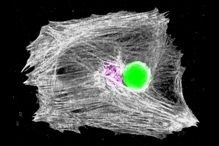
Image caption (top): Microscopy image of a heart muscle cell with a tiny embedded laser emitting bright green light. The white ribbons arching through the cell are part of the machinery that allows heart cells to contract. Analysing the colour of the light emitted by embedded lasers allowed observation of the workings of this machinery in an unprecedented manner, with important future implications for understanding heart disease.
The paper, 'Monitoring contractility in cardiac tissue with cellular resolution using biointegrated microlasers' by Marcel Schubert, Lewis Woolfson, Isla R M Barnard, Amy M Dorward, Becky Casement, Andrew Morton, Gavin B Robertson, Paul L Appleton, Gareth B Miles, Carl S Tucker, Samantha J Pitt and Malte C Gather is published in Nature Photonics and available online.
DOI: 10.1038/s41566-020-0631-z


