
Chlamydia organise their genetic material with the help of lipids. This is shown by a new study from the University of Würzburg. This unexpected alliance could change our current understanding of how bacterial genetic material is organised.
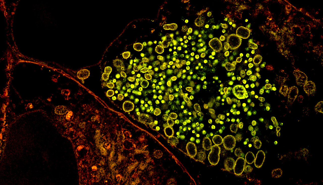
The bacterium Chlamydia trachomatis is one of the most common causative agents of sexually transmitted infections worldwide. Biologically, it is regarded as a highly specialised 'nutrient thief': as chlamydia have lost the ability to reproduce independently outside a host cell, they hijack human metabolic products - particularly sphingolipids - for their own development. Within the host cell, they create a sheltered environment, known as an inclusion. These bacterial colonies often grow to such a massive size that they even exceed the size of the human host cell's nucleus.
The life cycle of these pathogens is characterised by extreme morphological change. They alternate between robust, infectious elementary bodies for their time outside the cell and active reticular bodies that multiply inside the host cells. To protect the genetic material during the infectious phase, the DNA within the tiny elementary bodies is packed extremely tightly into a nucleoid.
Too small for a light microscope
For a long time, investigating this internal structure posed a strategic challenge for science: measuring just 200 to 300 nanometres in size, the elementary bodies lie below the physical resolution limit of conventional light microscopes. This circumstance even led to chlamydia being mistakenly regarded as viruses until the 1960s. New findings from Würzburg are now unravelling the mystery of this compact architecture.
The study, now published in the journal Nature Communications , was carried out by a team led by Professor Thomas Rudel, Head of the Chair of Microbiology at the University of Würzburg; the co-first authors are Marcel Rühling and Fabienne Wagner.
Lipids as the architects of the genome
In their study, the research team demonstrates that not only proteins but also lipids play a crucial role in the organisation of the bacterial genome. In particular, the sphingolipid sphingomyelin is incorporated directly into the highly compacted nucleoid. The spatial correlation is striking: around 90 per cent of the nucleoid in the infectious form is occupied by these lipids.
An overview of the study's key findings:
- Surprising localisation: Sphingolipids are found concentrated within the nucleoid of the elementary bodies - a location where these membrane components had not previously been suspected to be present.
- Earliest sign of transformation: The separation of lipids and DNA is one of the first measurable steps when the bacterium transitions to its active form after entering the host cell.
- Efficient packaging: The researchers speculate that the use of lipids for DNA compression is likely an energy-efficient strategy that partially replaces the resource-intensive synthesis of specialised proteins.
"To our surprise, in our experiments we observed the highly dynamic incorporation and removal of a sphingomyelin derivative in bacterial nucleoids during the developmental cycle," explains Thomas Rudel. This observation was only made possible by a technical combination that produces images with unprecedented levels of detail.
A trick for nanoscale insights
To overcome the physical limitations of optics, the team used expansion microscopy (ExM). The trick here is that, instead of zooming in with a better microscope, you simply make the sample itself larger. To do this, the biological sample is embedded in a swellable hydrogel which, when water is added, expands along with the sample to up to eight times its original size. Details that previously blurred into a single pixel thus become visible even with conventional light microscopes.
Images from a cryo-electron tomograph confirmed the observations made in this way. Whilst expansion microscopy allows specific molecules such as DNA and lipids to be colour-coded and identified, cryo-electron tomography provides high-resolution images of the bacteria in a flash-frozen state. It was only this combination of techniques that proved that specific membrane stacks form a structural bridge between the inner envelope and the DNA core. This combination of methods sets a new standard for the investigation of the smallest cellular structures.
New avenue for medical research
According to the scientists involved, the findings fundamentally alter our understanding of bacterial development. Lipid metabolism is far more than just a supplier for the cell envelope; it acts as a central regulator that controls access to the genes. The controlled release of DNA from its lipid packaging marks the starting point of the infection.
This knowledge offers a new strategic target for medicine: if this 'lipid-DNA bridge' could be specifically disrupted, the infection could be nipped in the bud. Nevertheless, questions remain. The exact molecular architecture at the interface between lipid molecules and genetic material remains partly unknown, given the current resolution limit of around 30 nanometres. Future studies must now clarify exactly how the binding occurs at the atomic level and whether this mechanism also exists in other specialised bacteria.
Original publication
Sphingolipids associate with the chlamydial nucleoid and mark developmental transitions in Chlamydia trachomatis. Marcel Rühling, Fabienne Wagner, Till Epprecht, Paul F. Köhling, Fabian Schumacher, Stefan Sachs, Louise Kersting, Julian Fink, Lina Girndt, Burkhard Kleuser, Markus Sauer, Jürgen Seibel, Gregor L. Weiss and Thomas Rudel. Nature Communications, DOI: 10.1038/s41467-026-77974-3


