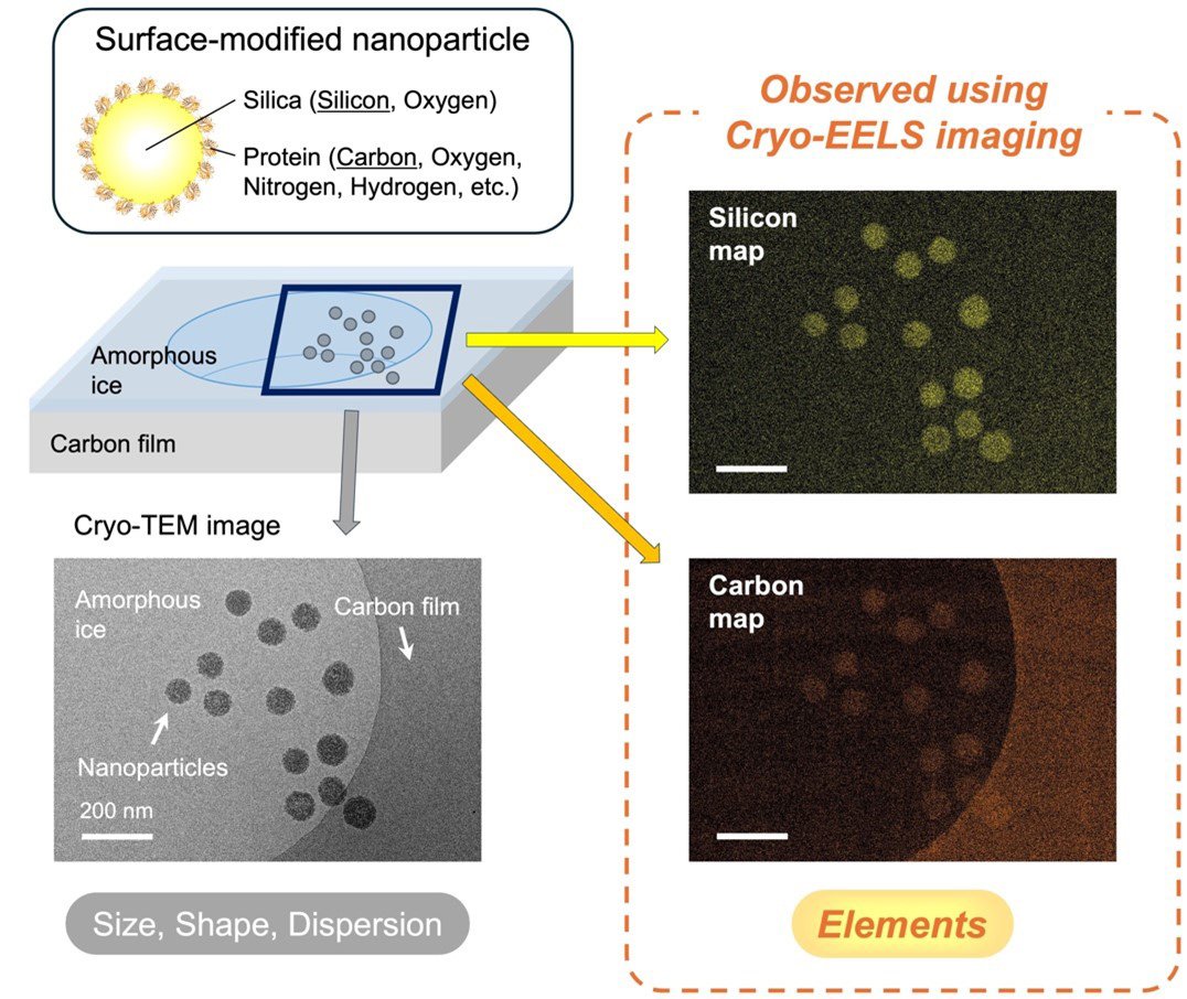
Cryo-transmission electron microscopy (cryo-TEM) allows us to observe samples in a preserved state that is close to their native form, making it a highly effective way to examine biological samples. This technique provides information on the size, shape, and dispersion of samples within a frozen solvent. However, there is another crucial piece of information that has not been accurately visualized in organic samples using this technique yet: elemental composition.
Energy-filtered TEM (EF-TEM) can determine the elemental composition by using electron energy loss spectroscopy (EELS), but it has its shortcomings. The conventional EELS/EF-TEM method can significantly damage the sample and usually exhibits drift - which results in a blurry image. Furthermore, conventional observations have primarily been applied to metals or large-area of samples, rather than to organic nano-materials.
To overcome these limitations, researchers at Tohoku University developed a novel elemental mapping technique using cryo-EELS/EF-TEM, enabling simultaneous visualization of both the structure and elemental distribution of nanomaterials in frozen solvents. This new approach allows high-resolution analysis of organic and bio-related materials.
The findings were published in Analytical Chemistry on July 31, 2025.
The team found that plasmon signals from ice strongly increased the background signal - an undesirable effect since they are focused on the organic sample and aren't interested in signals from the ice itself. To remedy this, the team developed a new imaging method combining the 3-window method for accurate background correction with drift compensation techniques. Additionally, a new program was created for the "ParallEM" electron microscope control system, allowing easy control of energy shift during imaging.
Using this technique, the team successfully visualized silicon distribution in silica nanoparticles within a frozen solvent, alongside their structure and dispersion. The smallest particle size detectable via elemental mapping was determined to be around 10 nm.
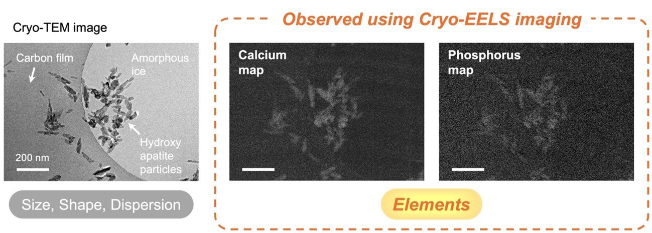
The method was also applied to hydroxyapatite particles, a major component of bones and teeth, revealing calcium and phosphorus distributions - key biological elements.
This technique is expected to enable advanced analysis of various materials, including biomaterials, medical materials, food, catalysts, and inks. As such, it can contribute to research across fields from life sciences to materials science.
- Publication Details:
Title: Low-Dose Elemental Mapping of Light Atoms in Liquid-phase Materials Using Cryo-EELS
Authors: Daisuke Unabara, Yohei K. Sato, Tasuku Hamaguchi, and Koji Yonekura
Journal: Analytical Chemistry


