Collaborative project developing new MRI scan for pregnant women
A collaborative artificial intelligence project between King's College London, Guy's and St Thomas' and Siemens Healthineers, is developing a new MRI scan on the UK's first MAGNETOM Free.Max scanner, for pregnant women to give more information to obstetricians about babies as close to birth as possible to optimally prepare for birth.
Study lead, Dr Jana Hutter, Senior Lecturer in the School of Biomedical Engineering & Imaging Sciences, has scanned more than 95 pregnant women for the Meerkat project which aims to develop a self-driving 'one-click' exam which also corrects for fetal movement in the womb.
Normally when an MRI is performed on a pregnant woman, there are different 'scanning steps' which are done one-by-one, but this is not adapted neither to the movement of the baby nor uterine practice contractions for example.
Dr Hutter is using AI to adapt in real-time depending on how the baby is moving. In this way, radiographers have almost like a 'rolling-feed' of the baby – in real-time – and can stop when needing to look at something in more detail.
For instance, if the fetus is moving a lot, the scanner will automatically change to a dynamic capture mode able to catch the baby's motion. And if the baby is not moving a lot, the time is used for high-resolution brain scans. Instead of always being suboptimal, the fetal scan thus adapts in real-time.
In-line with other ongoing research in fetal MRI at St Thomas' Hospital, the team are looking at a wide range of pregnancy complications such as pre-eclampsia and preterm birth as well as any clinical referral eg for anomalies in the babies' brains.
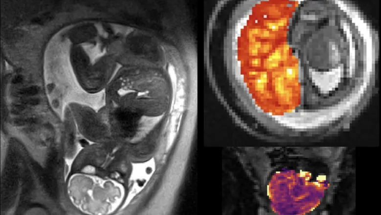
Through the examination that we have set-up as part of the study we see that we can get very reliable data from the placenta and the fetal brain. If we repeat the scan on different days, it is very reliable, robust and can be obtained very quickly on the low-field scanner. The researchers aim to ensure that they know how the pregnancy is progressing and what information the clinicians need to monitor them optimally.– Dr Jana Hutter, Senior Lecturer in the School of Biomedical Engineering & Imaging Sciences
This project requires very close collaboration with many experts, obstetricians to radiographers, midwives, radiologists as well as technical experts from Siemens Healthineers.
While 1.5Tesla or 3Tesla would be the normal magnetic field strength for an MRI system, which does yield similar information, the researchers have found using the lower magnetic field strength provides very rich information. Questions we are trying to address next are: will the placenta support the baby for the full pregnancy? Is it safe for the woman to have a vaginal birth?
The lower-field strength is particularly relevant in high-risk populations, such as for women with chronic hypertension, diabetes and bigger women. Due to the design of this scanner, there is a wider opening, meaning scans can more easily be performed on obese patients allowing for higher quality scans when patients have a high percentage of abdominal fat.
There are also impressive opportunities for pregnant women who are late in gestation, as radiographers can get all this information very close to birth.
Dr Raphael Tomi-Tricot, Senior MR Collaborations Scientist at Siemens Healthineers and visiting research fellow at the School of Biomedical Engineering & Imaging Sciences said the Perinatal imaging department at King's College London has a long-standing history of successful studies and investigations on fetal MRI, with very innovative ideas.
"Setting up this new set of developments on the MAGNETOM Free.Max scanner in such a short time is a real feat and it highlights the quality of the research there, with the well-being of the patient always at heart."
Thanks to the wider bore of the scanner, researchers can target a more vulnerable population, be it obese pregnant women or claustrophobic patients that would otherwise not have had access to MRI. It is also an example of the extremely rich collaboration between King's and Siemens Healthineers at St Thomas' Hospital, where we could put together expertise from both sides to harness the specificities of fetal scanning with innovative MRI sequences to further push the capabilities of this new scanner.– Dr Raphael Tomi-Tricot, Senior MR Collaborations Scientist at Siemens Healthineers
With the involvement of clinicians, the researchers are now looking at new cohorts, such as those with Crohn's disease, women with hyperemesis gravidarum (extreme vomiting during pregnancy), and other populations which are less reachable, as well as other higher-risk groups that can benefit from the accessible and shorter scan.


