Using total-body PET imaging to get a better understanding of long COVID disease is the goal of a new project at the University of California, Davis, in collaboration with UC San Francisco. The project is funded by a grant of $3.2 million over four years from the National Institute of Allergy and Infectious Diseases, part of the National Institutes of Health.
About 1 in 10 COVID-19 survivors develop a range of long COVID symptoms that can last from months to years. How and why these symptoms develop isn't completely known, but they have been linked to activated immune T cells getting into organs and tissues. Researchers also have linked long COVID to damage to the inner lining of blood vessels. These events can be related, because blood vessels become leaky when T cells are activated nearby, but may also be coincidental because leaky blood vessels allow more immune cells to leave the blood and enter tissues.

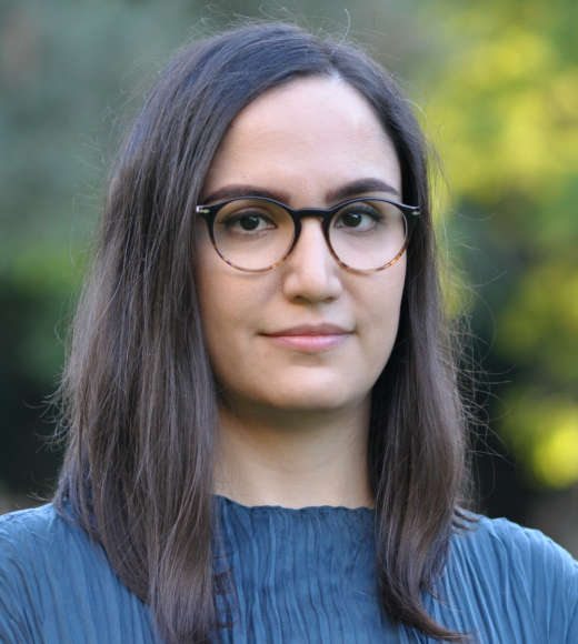
Negar Omidvari, assistant project scientist at the UC Davis Department of Biomedical Engineering and principal investigator on the grant, will use total-body positron emission tomography (PET) technology, originally developed by Professors Simon Cherry and Ramsey Badawi at UC Davis, and kinetic modeling to look at both processes simultaneously in patients with long COVID.
Imaging the entire body
PET imaging usually uses short-lived radioactive tracers to measure metabolic activity inside the body. Conventional PET can only look at a single organ or a section of the body at a time. The uEXPLORER PET scanner developed at UC Davis can image the entire body at the same time, giving a much more detailed picture of what is going on in the body.
Omidvari will collaborate with CellSight Technologies Inc. of San Francisco to use a tracer called 18F-AraG, which specifically tags activated T cells. Using dynamic total-body PET imaging and sophisticated modeling, she aims to see how activated T cells collect in different organs at different times, where blood vessel damage is occurring, and whether these processes are related to each other.
"If we can separate the vascular damage from the presence of activated T cells in tissue, we should be able to get a much better picture of what's going on," Omidvari said.
The team will also check blood samples for markers for inflammation and immune activation that correlate with PET imaging data.
The study will work with patients from UCSF's long COVID program (LIINC - Long-term Impact of Infection with Novel Coronavirus) who will be scanned at baseline, four and eight months. People who have fully recovered from COVID-19 and have no remaining symptoms will be scanned as controls.


