The Human BioMolecular Atlas Program has brought together 18 research teams, including collaborators from Vanderbilt, Harvard, CalTech, Stanford and others around the world, to build a comprehensive, three-dimensional molecular atlas of the human body.
On Sept. 1, HuBMAP released its inaugural data of several organs, including that of the kidney, mapped out by Jeffrey Spraggins, research assistant professor in biochemistry and director of BIOMIC and Richard Caprioli, Stanford Moore Chair in biochemistry and director of the Mass Spectrometry Research Center.
The BIOmolecular Multimodal Imaging Center, the NIH-funded HuBMAP tissue mapping center at Vanderbilt University, has been focused on developing an atlas for the human kidney for two years. BIOMIC includes experimentalists from Vanderbilt, clinicians from Vanderbilt University Medical Center and data scientists from Delft Technical University in the Netherlands.
"By working together in this multi-institutional, multidisciplinary environment, we can answer some of the biggest questions in medicine," Spraggins said. "It has been nice to interact so closely with peers from other institutions. We have exposed one another to new technologies and ways of thinking, which is driving innovation and enabling this immense human mapping project."
According to a release, HuBMAP's tools and maps are openly available for researchers to accelerate understanding of the relationships between cell and tissue organization and function. The consortium, managed by a trans-National Institutes of Health (NIH) working group of staff from the NIH Common Fund, National Heart, Lung, and Blood Institute, National Institute of Biomedical Imaging and Bioengineering and the National Institute of Diabetes and Digestive and Kidney Diseases, has released its first datasets on the heart, kidney, large intestine, lymph nodes, small intestine, spleen and thymus.
Being able to accurately visualize how the human body's trillions of cells interact, connect and arrange into tissues will provide greater understanding of human health. The use of all these data has multiple practical applications, including visualizing and explaining details of tissues and organs to patients or students, enabling software development of novel solutions, computational understanding of the differences between healthy and diseased cells, and precision drug development. HuBMAP has brought together universities-each with their own specialized technologies to generate images-and bioinformatics experts to integrate the data to bring as much information as possible to light.
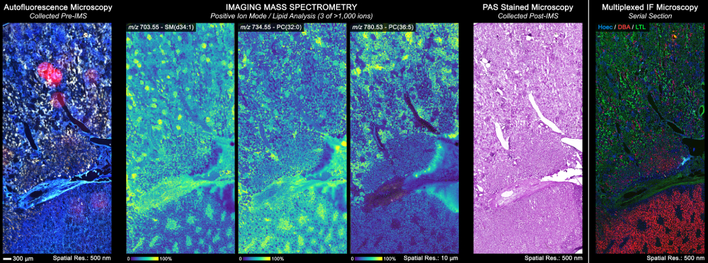
Caprioli developed imaging mass spectrometry technology, a system to map molecular distribution within tissues which was used in this project. in cancer tissue). Its combined deployment with other types of assays, including microscopy and MR/CT imaging, is unique among the research teams. "In a decade, we'll have the equivalent of 'Google Earth' for the human body," Caprioli said. "It has been a pleasure to use imaging mass spectrometry for such a significant project. We're using this tool to help scientists to gain a clear picture of molecular signatures and to unravel the complexity of human disease."
The kidney is the focus of a swath of diseases, including diabetes, acute kidney injury and chronic kidney disease, which is a focus HuBMAP kidney project collaborators Mark deCaestecker, Raymond Harris and Agnes Fogo. A critical component of kidney disease treatment is the understanding of its molecular progression. The Vanderbilt team posits that the HuBMAP atlas will enable physicians to look at organs where disease originates to more deeply understand its development.
The molecular and spatial atlas is still under construction, and the team at Vanderbilt recently received notice that HuBMAP will fund additional tissue mapping work of the pancreas and the eye over the next four years, to be led by Drs. Spraggins and Caprioli in conjunction with Kevin Schey, professor of biochemistry, ophthalmology and visual sciences, and Al Powers, Joe C. Davis Chair in Biomedical Science, Medicine, and Molecular Physiology and Biophysics.
BIOMIC is funded by the National Institutes of Health Common Fund grant U54DK120058 and U54EY032442.


