Too much communication in the Autistic Brain
An international research collaboration has developed a VR*1 imaging system that can measure a wide range of neural activity in the cortices of mice during active behavior. This enabled them to illuminate the abnormalities in cortical functional network*2 dynamics that are found in autism*3 model mice. Using machine learning*4, they were also able to highly accurately distinguish between autism model mice and wild-type mice based on the cortical functional network patterns when the mice start or stop running. The research group was led by Professor Toru Takumi and Assistant Professor Nobuhiro Nakai (both of the Department of Physiology, Kobe University Graduate School of Medicine), and Masaaki Sato, (Lecturer, Department of Pharmacology, Graduate School of Medicine, Hokkaido University). Professor Takumi is also a Visiting Senior Scientist at RIKEN Center for Biosystems Dynamics Research.
Future research on functional brain network dynamics in autism is expected to lead to the development of new biomarkers for autism diagnosis.
The results of this research was published in Cell Reports on March 28 at 11am (ET).
Main points
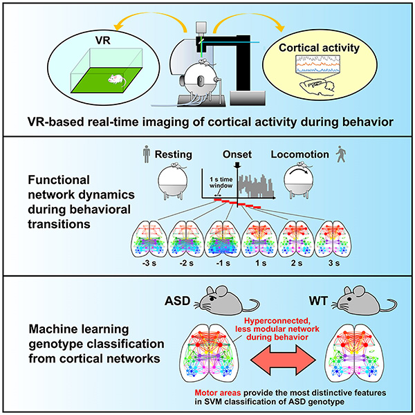
- The researchers developed a VR imaging system that can measure a wide range of cortical activity from mice in action.
- Mice models of autism have dense cortical functional networks and reduced modularity*5 after motor initiation.
- Machine learning can highly accurately identify autism model mice from their cortical functional network patterns.
Research Background
Autism (autism spectrum disorder) is a neurodevelopmental disorder with many unexplored aspects, characterized by poor social communication, intense preoccupation with certain things, and repetitive behaviors. The number of autistic individuals is markedly increasing, which is considered to be a significant social issue. Even now, autism diagnosis is based on behavioral characteristics, which is far from a quantitative perspective, and there is great demand for the discovery of a new biomarker.
In recent years, research has been conducted to identify functional brain abnormalities unique to autistic individuals. Resting-state fMRI*6 studies suggest that the density of functional brain networks increases in young autistic individuals and decreases in adults (Ref. 1). However, these changes vary widely from individual to individual. As the analysis was conducted when the participants were in a resting state, it was unclear how abnormalities in functional brain networks affect behavior.
Genetics contribute significantly to autism, and genomic abnormalities such as copy number variations (CNV)*7 are thought to be involved in neuropathology. Recently, animals (mainly mice) modeling human genomic aberrations are often used to elucidate the neuropathology of autism. In this study, the researchers developed a VR imaging system that can measure the brain activity of autism model mice in real-time during active behavior. By investigating brain functional network dynamics, the research group aimed to clarify autism-specific phenomena in the brain during behavior.
Results
First, a VR imaging system was constructed (Fig. 1A). A mouse with its head fixed in place is put on a treadmill and shown an image of a virtual space projected on a screen. The virtual space was prepared so that it reproduced the field used for mouse behavioral experiments. The motion of the treadmill is reflected in the video images, allowing the mice to freely explore the virtual space (Fig. 1B). Alongside behavioral measurements such as locomotion, transcranial calcium imaging*8 was performed simultaneously so that a wide range of functional area activity in the cerebral cortex could be measured in real time (Fig. 1C-E). For this purpose, the researchers used transgenic mice that express calcium sensor protein (GCaMP)*9 in their neurons. In addition, they established a method for analyzing cortical functional network dynamics. They calculated correlations between functional areas from one-second neural activity data obtained via calcium imaging, and visualized the functional network using graph theory (Fig. 1E).
The researchers analyzed the 3 second time windows before and after when the mouse spontaneously started or stopped moving on the treadmill (locomotion) and examined the network characteristics in each time window. The results revealed that the network structure changes with the onset of locomotion and that modularity increases (Fig. 2). It was also found that the network structure returns to the resting state when locomotion is stopped. Thus, they succeeded in visualizing the network dynamics during the switch from rest to locomotion and from locomotion to rest.


