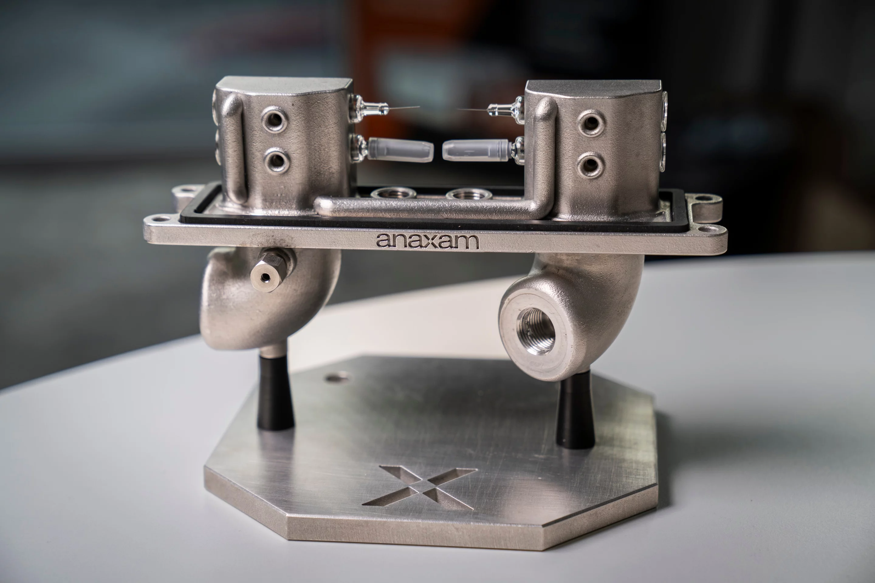
Researchers at the Paul Scherrer Institute PSI and the ANAXAM technology transfer center have used neutron and synchrotron imaging to determine the conditions under which pre-filled syringes become blocked.
(Photo: Paul Scherrer Institute/Mahir Dzambegovic)

(Photo: Paul Scherrer Institute/Mahir Dzambegovic)
(Photo: Paul Scherrer Institute/Mahir Dzambegovic)
In very rare cases, the needles of prefilled syringes may become blocked. This can have potentially detrimental consequences for patients if their medication does not enter the body or the dosage is too low. A team from the Paul Scherrer Institute PSI, led by the ANAXAM technology transfer center in collaboration with MSD (a tradename of Merck & Co., Inc., Rahway, N.J., USA), has now been able to take a detailed look inside such needles. The researchers were able to determine the possible causes of the blockages and create the conditions to prevent them in future. The key was a combination of imaging techniques at the Swiss Light Source SLS and the neutron source SINQ. Both are located in close proximity to each other on the PSI site - an ensemble that is unique in the world. The result is a first, say chemical engineer Vladimir Novak and physicist and CEO of ANAXAM Christian Grünzweig: "We have succeeded in taking the most detailed look yet at the inside of a needle."
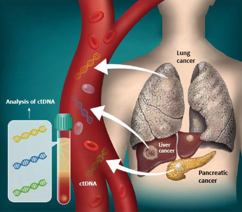
Pre-filled syringes (PFS) are increasingly used for cancer treatment, for example using monoclonal antibodies. However, in a small number of cases the needles of the pre-filled syringes clog. The risk of this has increased since the therapeutic agents, which consist of proteins and antibodies, have become more concentrated and therefore more viscous, offering greater resistance to fluid flow. No reliable figures are available on the average frequency of a blockage. Researchers are discussing fluctuating pressure or temperature conditions as possible causes, for example while transporting batches.
Pressure and temperature fluctuations as possible causes
The team led by chemical engineer Vladimir Novak, head of the project group and researcher at ANAXAM, investigated this hypothesis in a series of measurements. For example, 31 PFS were exposed to changes in temperature between 5 and 40 degrees and changes in pressure between 550 and 1010 millibars. Such conditions typically also occur during flights when transporting medical products. The team then examined the needles using both synchrotron X-ray and neutron beams.
Both imaging methods have certain advantages. Neutrons can penetrate metals better, but are deflected by hydrogen atoms, which occur in liquids, and thus produce a clear contrast. "Neutron radiography allows the liquid inside the needle to be visualized in 2D, whereby both the ambient pressure and temperature can be varied while measuring the syringes," explains David Mannes, from Laboratory for Neutron Scattering and Imaging of PSI.
Conventional X-ray computed tomography is normally unable to visualize low-density liquids inside stainless steel in such detail, because the transmitted light beam is not attenuated enough. However, this can be achieved using partially coherent beams, like those available at PSI's SLS.
Synchrotron X-ray tomography offers high-resolution 3D imaging for studying detailed interfaces between air and liquid inside a needle. "This new application of the technique represents the first attempt in the scientific community to investigate the problem of needle clogging using synchrotron X-ray tomography", explains Margie Olbinado, Industrial Liaison Scientist from PSI.
The measurements showed clearly that fluctuations in the ambient conditions led to deposits on the metal of the needle. Both pressure and temperature fluctuations drive liquid into the needle, where air bubbles may also form. When the liquid dries, it leaves behind plugs or deposits inside the needle. Manufacturers, distributors, and users should therefore ensure an uninterrupted cold chain and constant pressure when transporting and storing the PFS. "The combination of neutron imaging and synchrotron X-ray tomography clearly demonstrates this," says Novak.
Text: Werner Siefer