What the research is about
The sweet aroma of sugar and freshly baked bread-sugars are a familiar part of our everyday life. Inside our bodies, however, sugars play far more important roles, supporting communication between cells and regulating immune responses. When sugars are linked together like a chain, they form structures called glycans, which are essential for many biological processes.
One challenge in studying glycans is that they are extremely flexible. The bonds between sugar units can rotate freely, causing the overall shape to change constantly. Because of this motion, it has been very difficult to capture the precise three-dimensional structures of glycans.
To overcome this problem, researchers have explored the idea of fixing sugars onto protein crystals and observing them using the protein as a scaffold. An ideal scaffold protein must firmly hold the target molecule and align it in a uniform orientation. If this can be achieved, even highly mobile sugars can be observed as if their motion were "frozen in time."
Among the proteins studied for this purpose, galectin-10 (Gal-10) has attracted particular attention. Gal-10 is a rare protein that naturally forms crystals inside the human body, making it seem as though it already possesses the ideal scaffold structure. However, reproducing this crystallization process in a test tube has proven far more difficult than expected. Researchers must prepare large amounts of highly pure Gal-10 and carefully adjust crystallization conditions-such as temperature, concentration, and solution properties. Because this process requires enormous time and effort, it has long remained unclear how everyday sugars are actually captured by Gal-10 at the molecular level.
Why this matters
A research team led by Professor Takafumi Ueno at Institute of Science Tokyo (Science Tokyo) has overcome this long-standing challenge. The key breakthrough was eliminating the need for time-consuming protein purification and weeks-long crystal growth. Instead, the team developed a method that produces proteins and crystallizes them at the same time in a test tube.
In this approach, no living cells are used. By simply mixing the necessary components, uniform protein crystals can be formed rapidly and reliably. Using this method, the team succeeded in producing stable Gal-10 crystals within just 24 hours. This innovative technique is known as the cell-free protein crystallization (CFPC) method.
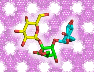
With this platform, the researchers achieved several world-first results. They determined the crystal structure of the trisaccharide melezitose-a sugar found in the sweet sap of trees such as poplars-bound to a protein scaffold. This achievement made it possible to clearly visualize the three-dimensional structure of a molecule composed of three linked sugars.
The team also succeeded in revealing how lactose, the sugar found in milk, binds to the natural (non-modified) form of Gal-10-something previously thought to be impossible. They showed that lactose binds in a different orientation than expected.
By combining crystallographic analysis with molecular simulations, the researchers were able to quantitatively explain how specific amino acids in the protein restrict the movement of sugars and stabilize their structures-another major advance of this work.
What's next
This technology has direct implications for drug discovery. Glycans often act as "molecular signposts" that viruses, bacteria, and cancer cells use to interact with the human body. Understanding how glycans bind to proteins at the molecular level can guide the design of new drugs and enable rapid screening of unknown molecular structures.
Trisaccharides such as raffinose are also attracting attention as prebiotics that promote the growth of beneficial gut bacteria. The structural insights obtained in this study will contribute to the development of functional foods and new approaches in healthcare and nutrition.
Comment from the researcher
Sugars were difficult to study because they are always moving. By using crystals as a molecular 'photo studio,' we were finally able to capture their shapes. We hope this approach will deepen our understanding of life at the molecular level and lead to applications that benefit society.
(Takafumi Ueno, Professor, School of Life Science and Technology, Institute of Science Tokyo)