PhD student Daniel Cromb, from the School of Biomedical Engineering & Imaging Science has been awarded $5,000 from the International Society for Magnetic Resonance in Medicine (ISMRM) as part of their Research Exchange Program.
Funding will enable Dr Cromb to travel to Utrecht, The Netherlands for several months to continue developing a long-standing collaboration with a team at the Wilhelmina Children's Hospital (WKZ) at the University Medical Centre (UMC) Utrecht who have similar research interests.
While in Utrecht, Dan will be working closely with PhD student and medical doctor, Dr. Maaike Nijman, Professor of neonatology, Prof. Manon Benders and Associate Professor, Dr Jannie Wijnen.
We will be working together using placental histology and fetal brain MRI data to try and answer the question: 'Is impaired in-utero brain development in congenital heart disease, identified on fetal MRI, associated with placental abnormalities?'– Dr Daniel Cromb, PhD Student
Supervised by Prof. Serena Counsell and Dr Jana Hutter, Cromb's research is investigating the relationship between placental and early brain development and how these are affected in the context of congenital heart disease.
Exploring the relationship between fetal brain volumes, fetal-to-neonatal brain growth and placental histopathology could provide insight into the mechanisms underlying the impaired brain development seen in CHD and help identify potential biomarkers for early detection and treatment of the disease.– Professor Serena Counsell, Perinatal Imaging & Health
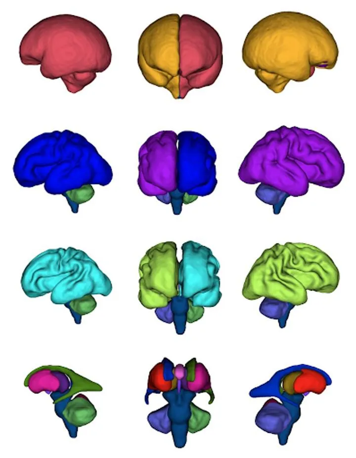
The team are using advanced Magnetic Resonance (MR) imaging techniques to acquire fetal brain, placental and neonatal brain data, along with placental histology and comprehensive clinical information. This will help them to better understand factors that lead to the altered brain development and adverse neurodevelopmental outcomes that are seen in some children with congenital heart disease.
The placenta is the interface between mother and fetus, providing the fetus with oxygen and nutrients, and plays a crucial role in organogenesis. Histological studies have identified abnormalities in placental structure and function in CHD and it is possible that these abnormalities are associated with impaired brain development. However, little is known about the role of the placenta in early brain development in individuals with CHD.