How are synapses formed, those points of contact that allow the transmission of information from one neuron to the other?
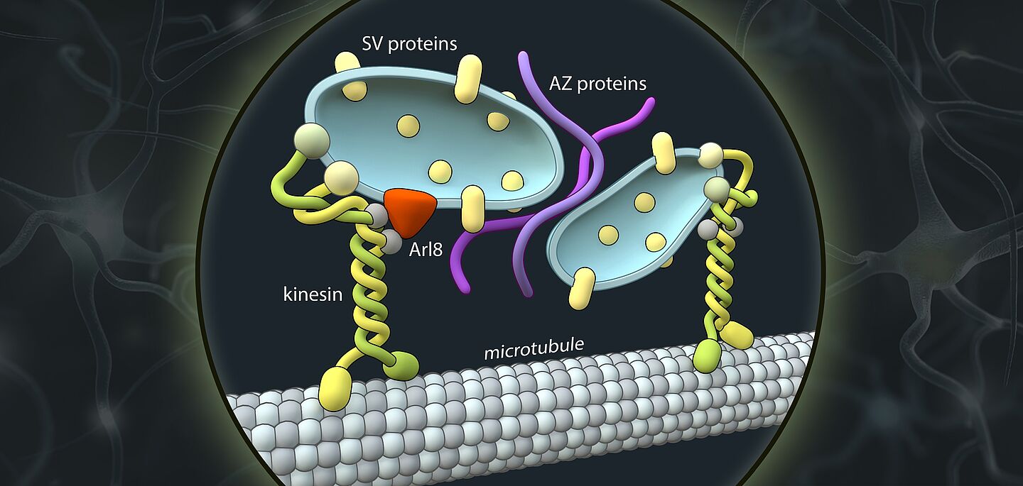
Schematic representation of axonal transport vesicles (blue) carrying presynaptic proteins (SV and AZ proteins). Kinesin motor proteins (KIF1A) attach these vesicles and carry them along the axons to the site of synapse formation. | Visualization: Barth van Rossum
Working with an international team, researchers from the Leibniz-Forschungsinstitut für Molekulare Pharmakologie (FMP) have now uncovered a crucial mechanism and elucidated the identity of the axonal transport vesicles that generates synapses. The findings provide an important basis for promoting the regeneration of nerve cells and counteracting the aging process in the future. The results have just been published in the journal Science.
Whether in the brain or in the muscles, wherever there are nerve cells, there are synapses. These contact points between neurons form the basis for the transmission of excitation, i.e. communication between neurons. As in any communication process, there is a sender and a receiver: Nerve cell processes called axons generate and transmit electrical signals thereby acting as signal senders. Synapses are points of contact between axonal nerve terminals (the pre-synapse) and post-synaptic neurons. At these synapses, the electrical impulse is converted into chemical messengers that are received and sensed by the post-synapses of the neighboring neuron. The messengers are released from special membrane sacs called synaptic vesicles.
As well as transmitting information, synapses can also store information. While the structure and function of synapses are comparably well understood, little is known about how they are formed.
A team from the Leibniz-Forschungsinstitut für Molekulare Pharmakologie (FMP)in Berlinhas now shed significant light on this mystery. Scientists from Charité-Universitätsmedizin, the Max Delbrück Center for Molecular Medicine (MDC) and the Universities of Leipzig, Chicago and Sheffield also contributed to this remarkable work.
Fluorescent protein reveals development of synaptic vesicles


