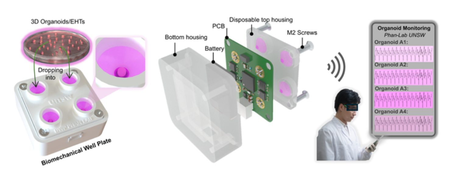
An international team, including the University of Tokyo, has created a sensor inspired by the lateral line in fish – their "sixth sense" organ – which measures the pulse of lab-grown 3D heart tissue (cardiac organoids). The device, called a biomechanical well plate, looks like a small white box containing four liquid-filled wells. When an organoid is placed in a well, each beat causes the liquid to bulge into an air cavity below, changing the air pressure. This change bends a cantilever sensor, which sends live data wirelessly to an app. The device is precise, reusable and less labor-intensive than some other methods. Due to its scalability, hundreds of tests could potentially be monitored simultaneously, with advantages for drug screening and future personalized medicine.
Cardiovascular research has undergone a revolution over the past 10 years, thanks to the development of 3D cardiac organoids. These lab-grown bundles of cells, while not full replications of a human heart (they are typically no bigger than 3 millimeters), still have enough detail that researchers can use them to study heart development, disease and the effects of new treatments. Before now, studies relied on flat 2D cell cultures or animal testing, both of which are limited in how accurately they can reproduce the behavior and responses of the human heart.
Small and complex, studying cardiac organoids can be challenging. Depending on the purpose, organoids may be directly grown on to sensors, limiting their reuse, or analyzed one by one via microscope, which takes time and makes simultaneous testing difficult. However, a cross-disciplinary and international team from Australia, the U.S. and Japan have now developed a device which could measure the pulse of hundreds of cardiac organoids all at once, ramping up the scale and speed of testing.

"Our device measures the pulse strength and rhythm of cardiac organoids using a biomechanical multi-well plate as the foundation. The design means that you can parallelize many measurements, testing different types and concentrations of treatments, while wirelessly receiving the data to see how the organoids respond in real time," explained Associate Professor Timothée Mouterde from the Graduate School of Engineering at the University of Tokyo.
One biomechanical well plate contains four liquid-filled wells with small holes at the bottom, air cavities beneath each well, and a delicate cantilever-based sensing system below. As an engineer specializing in fluid dynamics and surface interfaces, Mouterde's role was to figure out how to make the delicate interface between the liquid, the air cavity and the sensor work.
"The challenge was that there is no direct contact between the liquid that holds the heart organoids and the sensor. Instead, we created a water interface which traps an air cavity below, and the only reason it does not flood the cavity is because of carefully managed surface tension which we first worked out through analytical computer models," explained Mouterde.
"It is a fine balance, as the liquid needs to be able to move into the air pocket without flooding it. The beating of the organoid deforms water into the cavity before bouncing back, causing pressure fluctuations which compress the air, activating the cantilever sensor below and enabling us to pick up the heartbeat."
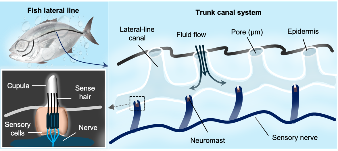
The device was inspired by a biological feature in fish called the lateral line, sometimes referred to as their "sixth sense." This organ runs along the body and water enters it through tiny pores, pushing against rows of small gelatinous caps called cupula. These cupula cover sensory hair cells, which translate changes in cupula movement into neural signals, indicating nearby prey, predators and other information about the surrounding environment.
"Because our device detects change in pressure, it is very good for measuring the fluctuations of a heartbeat and how it may change – becoming faster, slower or more irregular – in response to drug treatments," said Mouterde. "It also has a key advantage over animal testing as we can directly test drug treatments on human tissue, opening the way for future more personalized drug therapies which consider a person's individual genetics. As an engineer rather than a biologist or pharmacologist, our research demonstrates the advantages of cross-disciplinary collaboration and what we can build together."


