The finalists of QUT's annual Research in Focus multimedia competition have again shone a creative light on the university's diverse research.
The winners of the 2022 contest will be announced this Thursday at a showcase at The Cube (4.30pm to 6pm) at Gardens Point campus, including the People's Choice award (voting via Facebook closes Tuesday at 11.59pm).
A judging panel sifted through this year's entries to shortlist the following 10 finalists.
2022 QUT Research in Focus finalists
Dusan Bojic – SpiroArtis Image: This artwork has been produced from the breath of an adolescent patient with respiratory disease, using SpiroArtis – the world's first art-based interactive health technology platform, founded by QUT Doctorate in Creative Industries student Dusan Bojic as a creative way to add incentive for lung volume testing.
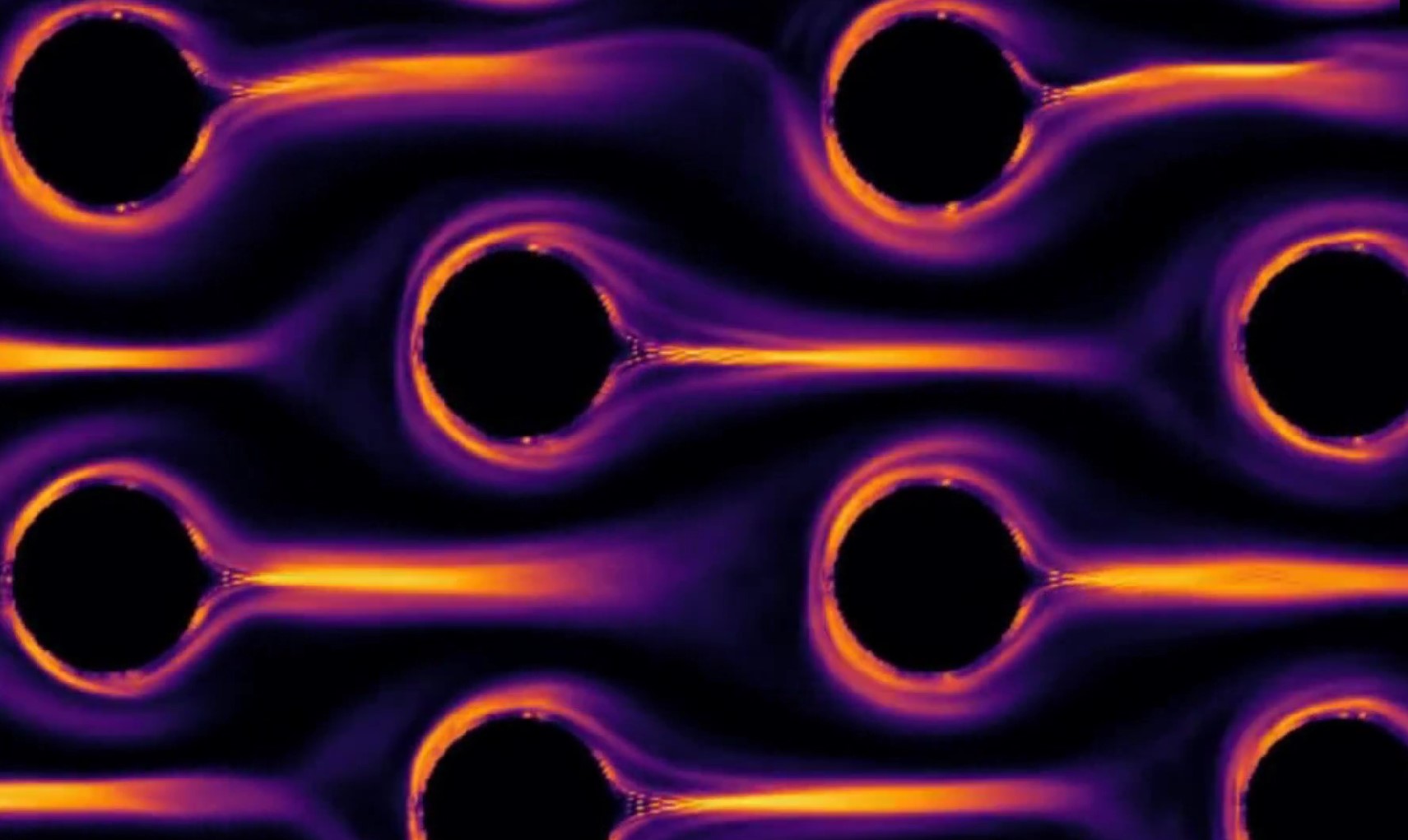
Vedad Dzanic – Hypnotic Dance of the Polymers: The animation shows the numerical simulation of viscoelastic fluid (a solvent fluid mixed with polymer additives) flowing past cylinder obstacles. When polymers stretch excessively, viscoelastic fluids transition into a chaotic state, coined elastic turbulence. (Click image below to view video.)
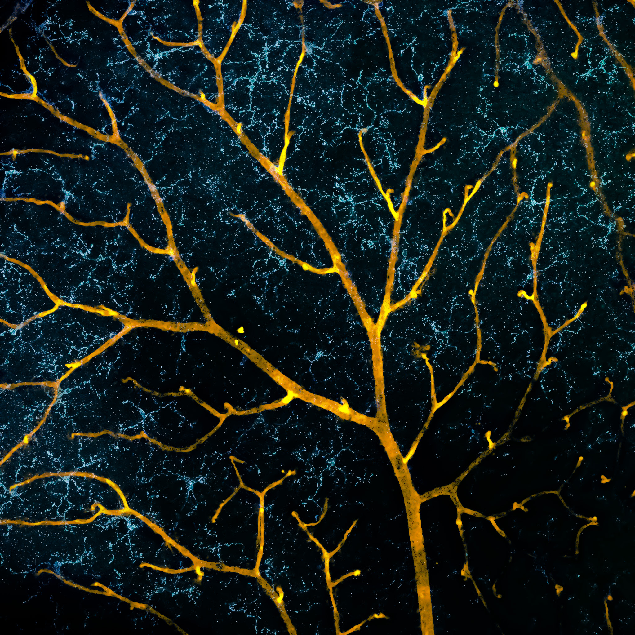
Fazeleh Etebar – Blossoms of the eye: This is a confocal fluorescence microscopy image of a mouse retina showing a layer at the back of eye. The colours represent the vasculature (gold) and microglia (blue).
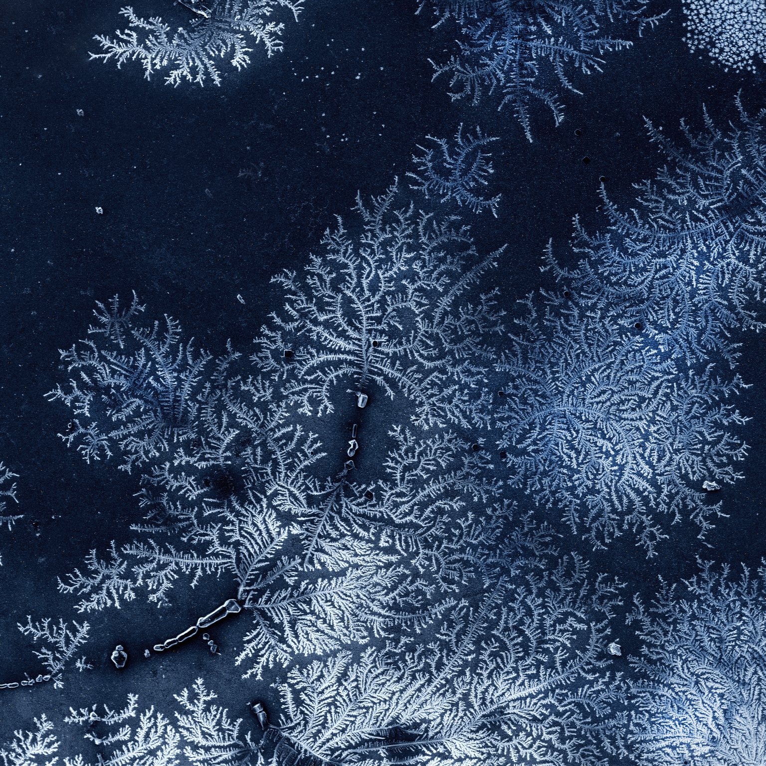
Konstantin Faershteyn – Frozen fractals all around: Rapid solidification of salt often results in a characteristic tree-like structure of crystals – dendritic structures – as this scanning electron microscopy image depicts. This multi-branching form is also typical for snowflakes or so-called "frozen fractals".
John Griffin – The Next Generation: This is a sorus of a fern at QUT's Gardens Point campus, showing the native fluorescence of the sample under the lasers of the microscope. The image demonstrates the capabilities of one of QUT's advanced imaging platforms.
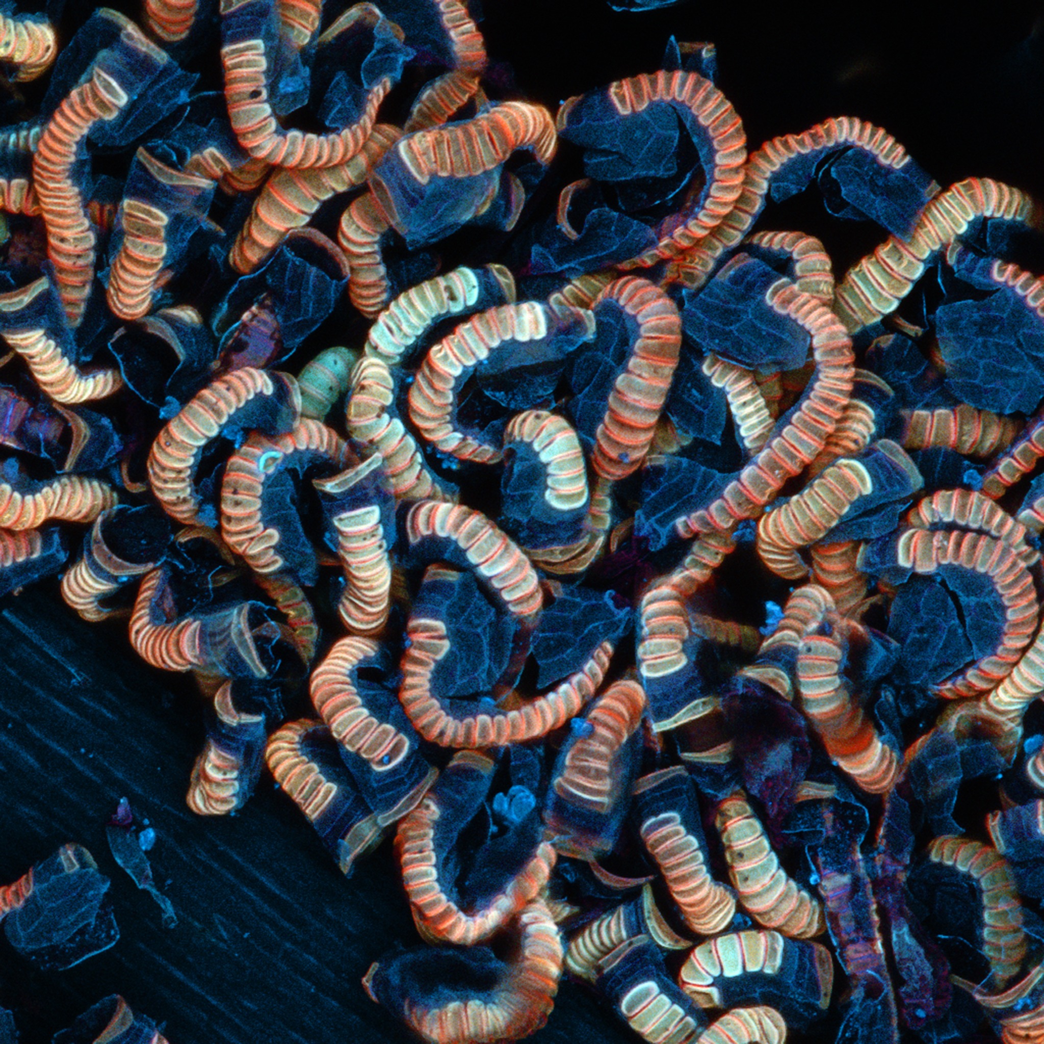
Lisa Kearney – Keeping It Together: This is a multi-element map of reef rock produced with the Tescan TIMA scanning electron microscope at QUT's Central Analytical Research Facility. The TIMA produces large-area maps that can be used to observe samples at varying scales. Like research students, coral reefs are often trying to keep it together, finding support in a unique community that surrounds them.
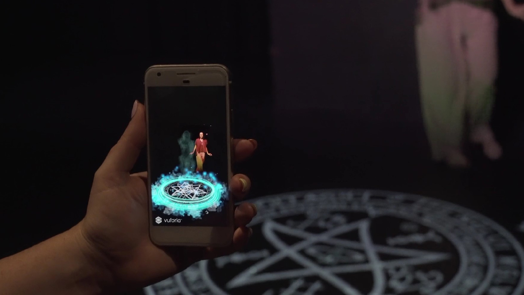
Kristen Maloney – Ghost Story: This is a cross-industry collaboration between performance and computer programming to create an innovative augmented performance. This new form of digital performance integrates live performance and augmented realty and is experienced via a smartphone app. (Click image below to view video.)
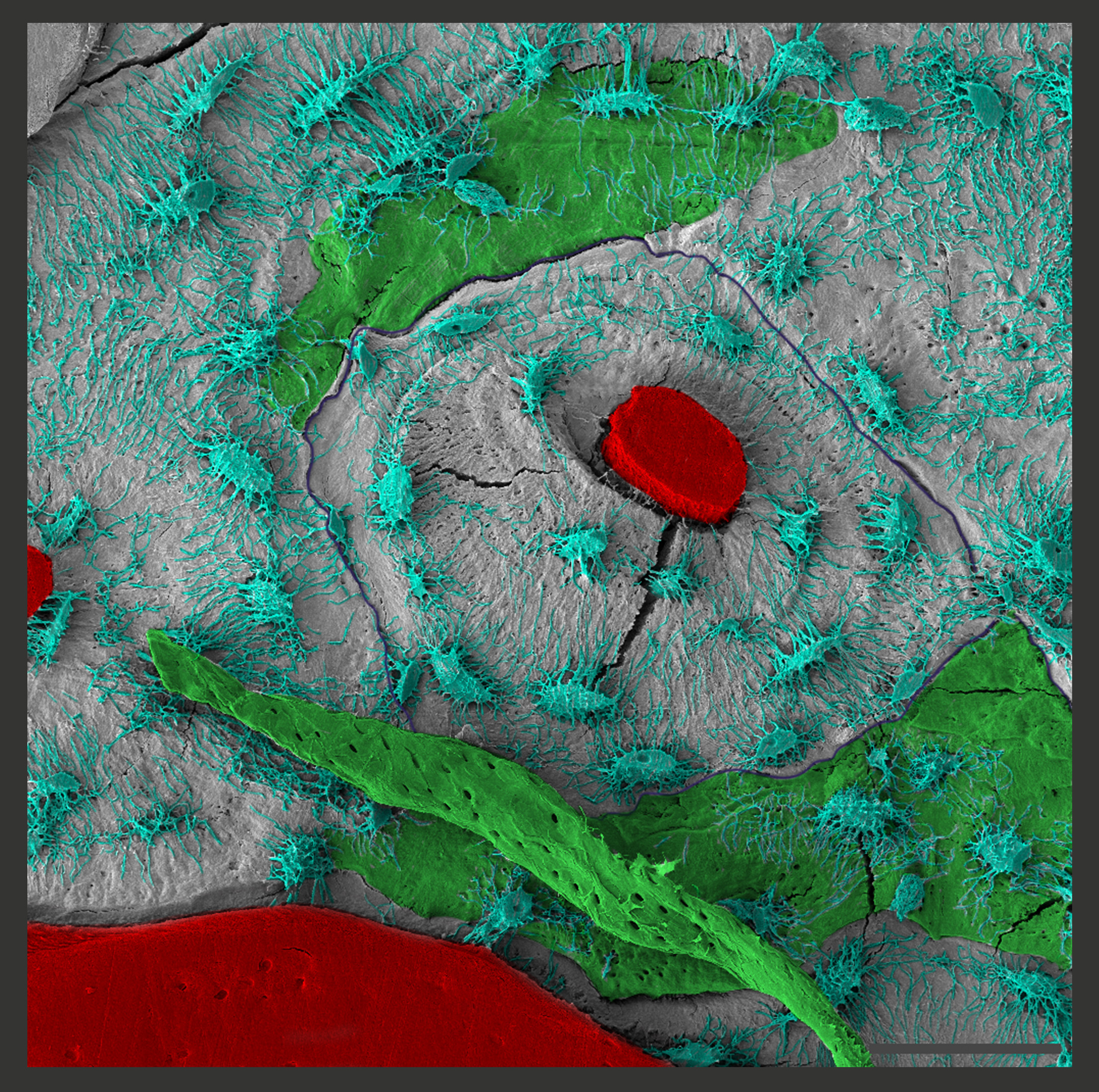
Flavia Medeiros Savi – Osteons in focus: This scanning electron microscopy image shows a 'region of interest' of a 3cm tibial bone defect, reconstructed with a medical grade polycaprolactone scaffold in combination with a corticoperiosteal flap. The image shows the new bone tissue being built, and the osteocyte network distributed around the haversian canal.
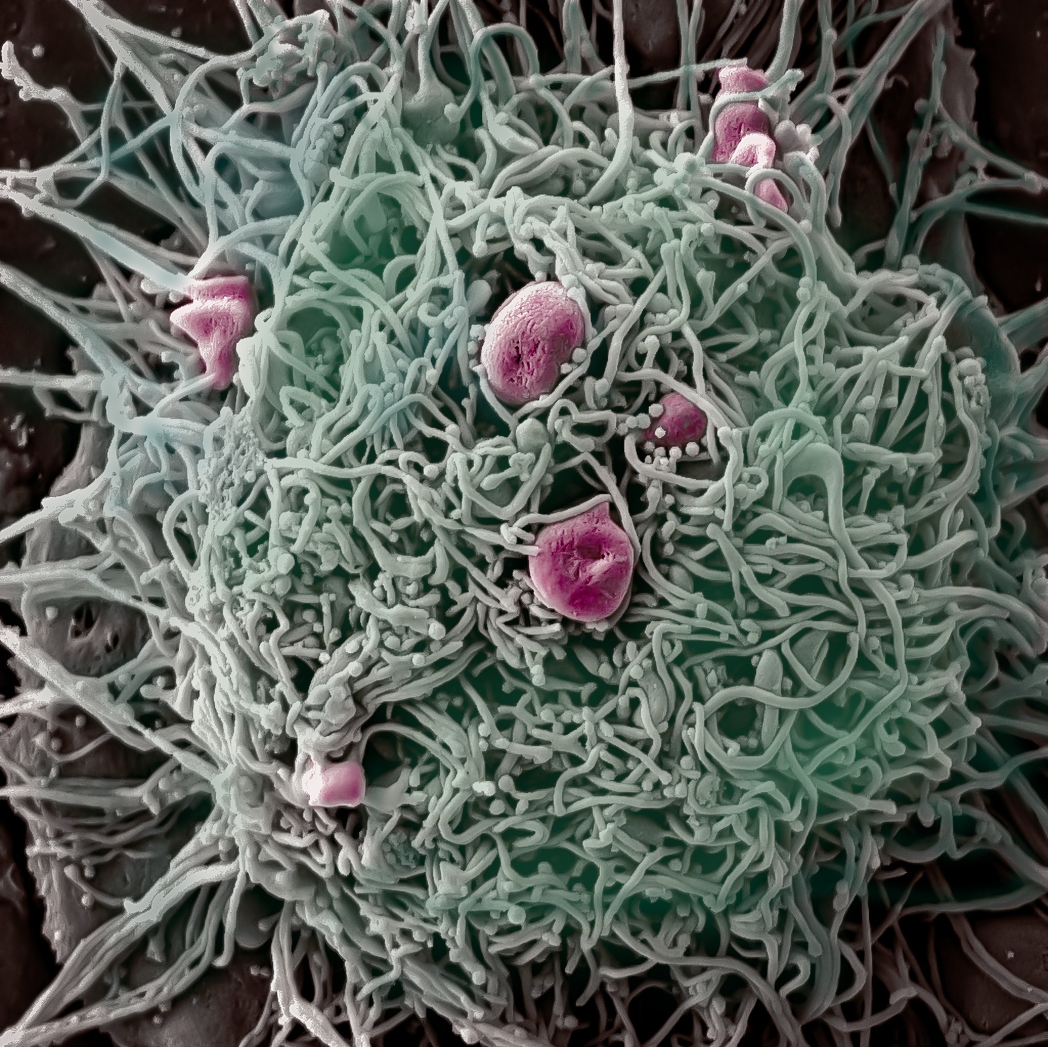
Jayanti Mendhi – Stay hungry, but don't stay foolish: Macrophages are a category of immune cells known as 'phagocytes, a word of Greek origin which means 'the cell that eats or devours'. This is an electron microscopy image showing bacteria (in pink) being engulfed by an activated macrophage cell. The aim of this research study was to understand how an antimicrobial coating which successfully killed bacteria impacted the ability of macrophages to eat up these bacteria.
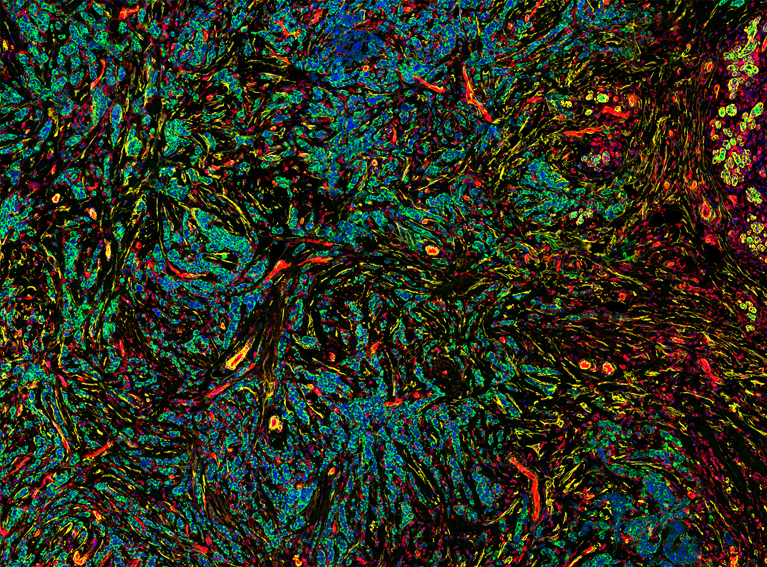
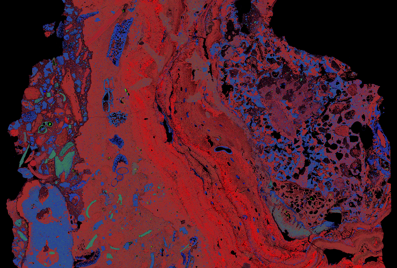
Akhilandeshwari Ravichandran – Illuminating Cancers: This GeoMx digital spatial profiler scan shows the tumor region of a triple negative breast cancer patient's tissue section, illuminated using fluorescent morphology markers – PanCK (green, cancer cells), DNA (blue, all cells), SMA (yellow) and CD31 (red, endothelial cells).