Scientists from the TSU Laboratory of Neurobiology, in the course of a series of experiments conducted using a model of ischemic stroke in rats, obtained new data on the processes that occur in the focal point of brain damage. This information can be the basis of a noninvasive diagnostic method that assesses the degree of inflammation, to predict the course of the disease and choose a treatment strategy for patients who have survived a "vascular catastrophe". The research results are published in the highly ranked International Journal of Molecular Sciences.
To study the formation of new neurons in ischemic stroke, a special method of labeling young neurons was created. TSU scientists, with colleagues from the KU Leuven (Belgium), designed a courier for transporting genetic material into brain cells - special vectors based on lentiviruses and adeno-associated viruses. A similar method of delivering genetic material is now being used in the development of some vaccines and drugs.
Genetic engineers inserted a ferritin protein gene and a special genetic sequence into the neutralized virus that helps to increase the production of ferritin only in young neurons. Young neurons that have accumulated ferritin-containing iron atoms can be seen using a special magnetic resonance imaging (MRI) protocol.
- When scanning the brains of animals in which ischemic stroke was simulated, we saw two areas with a specific change in the MRI signal, indicating the presence of a large number of cells containing ferritin,- says Marina Khodanovich, head of the TSU Laboratory of Neurobiology at the Research Institute of Biology and Biophysics. - The signal was recorded in the nonischemic zone, where the active production of young neurons usually begins after a stroke. This did not come as a surprise to us, but the presence of the same signal in the stroke focus was unexpected.
A subsequent study of brain sections showed that such a signal was given by macrophages - cells of the immune system that are also called "big devourers" (from ancient Greek μακρός - large, and φάγος - devourer). Macrophages are capable of actively trapping and digesting bacteria, particles foreign or toxic to the body, and fragments of dead cells. After a cerebral stroke, macrophages migrate to the ischemic focus, where they absorb not only the destroyed nerve tissue but also iron-rich erythrocytes, which makes them visible on MRI.
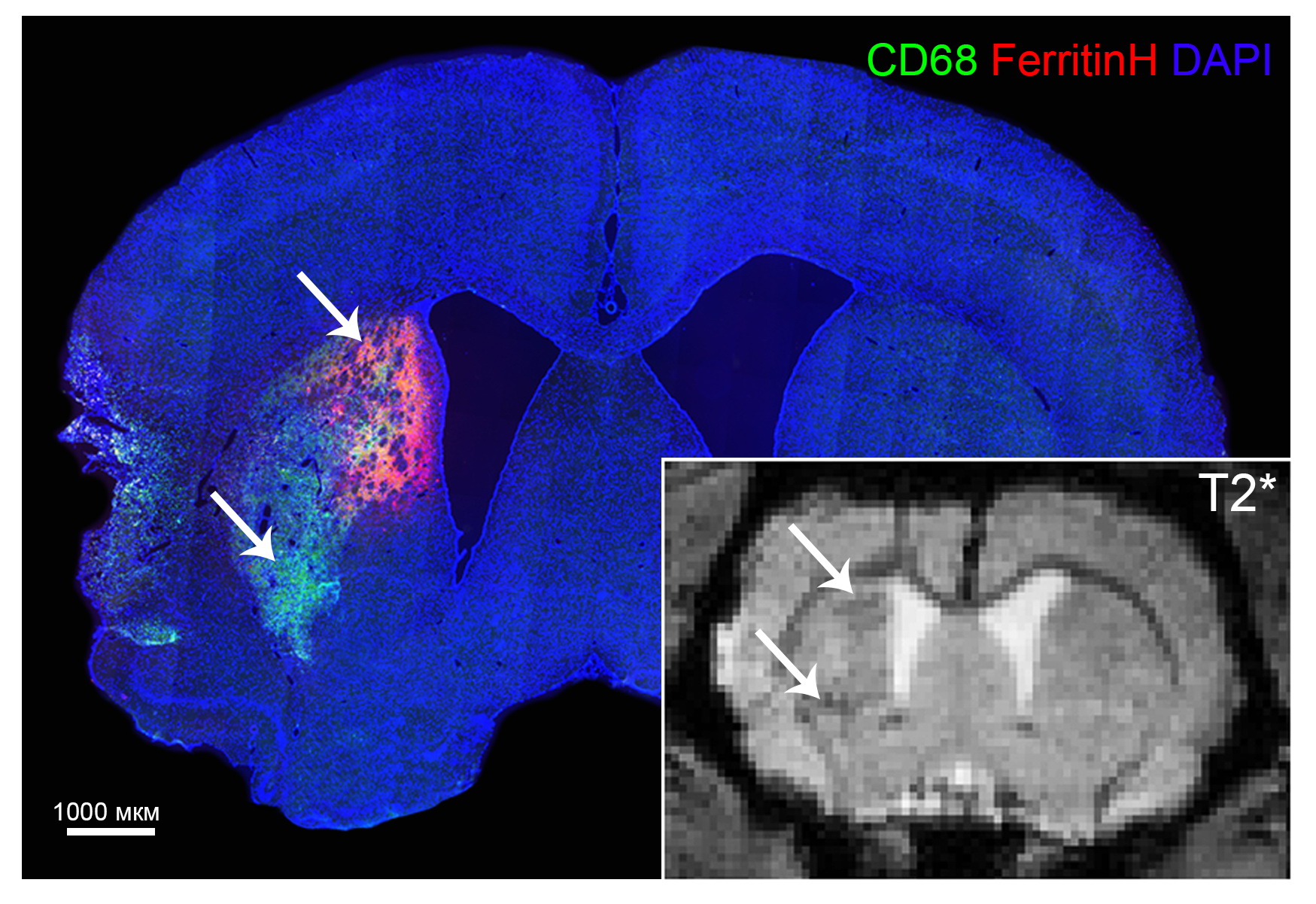
According to TSU neurobiologists, MRI observation of macrophage clusters can be used to create a new diagnostic approach that will be useful to clinicians by making it possible to assess the intensity of inflammation in the stroke focus, obtain more information about the patient's condition, more accurately predict the course of the disease, and select drug therapy.
Now the main challenge for neuroscientists is to find a way to differentiate signals from macrophages and new neurons with genetic marks. For this, it is necessary to continue the research begun to obtain additional fundamental knowledge about the behavior of macrophages and new neurons in the focus of ischemic lesion.


