
Instead of treating movement over time as a limitation, the system uses those changes to show more detail than a conventional camera could, opening up new possibilities for studying cells, and (even in the summer) snowflakes
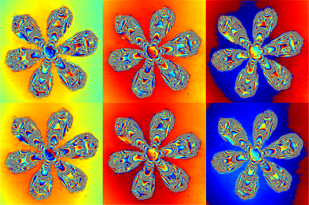
Six moments of a melting snowflake, captured with the lensless chip. Each frame is a phase image in which color represents the thickness of the ice. The densely packed color fringes are contours of equal thickness that trace the 3D relief of the crystal's arms. The color washing across the background is a nanometer-thin film of meltwater on the substrate, a change far too subtle for any ordinary camera to see. (Guoan Zheng/UConn)
For the first time ever, researchers can witness nanometer-scale thickness changes across a melting snowflake, with a compact, lensless chip that captures gigapixel video at 30 frames per second across centimeter-scale fields.
Snowflake imaging has fascinated scientists and the public since the first snowflake images were captured in 1885 by Wilson Bentley, establishing that no two snowflakes are identical.
The new system reveals what Bentley could never capture. The accompanying images follow a single snowflake as it melts. What looks like the petals of a flower is actually a map of the ice's thickness: each color band marks a change in height, and the shifting background traces a nanometer-thin layer of meltwater spreading on the substrate, details no previous instrument could record across a centimeter-scale field.
The study, "Video-rate Gigapixel Ptychography Via Space-time Neural Field Representations," was published in Nature Communications in June, and also points to a better way of monitoring living cells.
"We're introducing a new way of imaging that lets us record very large areas at an extremely high resolution and video speed," says biomedical engineering professor Guoan Zheng. "We've accomplished this by combining specially designed optics with AI."
Instead of treating movement over time as a limitation, the system uses those changes to reveal more detail than a conventional camera could, opening up new possibilities for studying cells, medical devices, and (even in the summer) snowflakes.
The work was an interdisciplinary UConn Engineering effort spanning biomedical engineering (Zheng), chemical and biomolecular engineering (Leslie Shor), the School of Mechanical, Aerospace and Manufacturing Engineering (Thanh Nguyen), and CLAS's molecular and cell biology (Daniel Gage). Co-first-authors Ruihai Wang, Qianhao Zhao, and Zhixuan Hong developed the space-time neural field framework, and the work was carried out with the Department of Energy's Pacific Northwest National Laboratory (Mary Lipton, Christopher Anderton, and Arunima Bhattacharjee) and David Brady of the University of Arizona.
Traditional cameras use a lens to focus light onto a sensor. With lensless ptychography, light passes through a specially designed surface before reaching the sensor. A computer knows exactly how the light was scrambled onto the surface, then mathematically reconstructs the image with more information than a normal camera could.
As the images are captured, the system uses a space-time neural field (a type of AI model), to learn the entire video as one continuous object rather than thousands of separate frames. By drawing on the correlations between frames, it lets every measurement do double duty: each one sharpens the spatial detail toward gigapixel scales, while keeping the full video frame rate.
The result is gigapixel video that records not just a flat picture but the phase of light, which converts directly into height. It resolves features finer than a thousandth of a millimeter, more than a hundred times thinner than a human hair, across a centimeter-scale area.
Beyond Snowflakes
Besides snowflakes, the imaging method can improve work across many scientific applications.
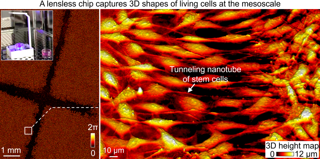
To show what the method offers biology, the team placed the chip inside an incubator and recorded living stem cells as they closed a scratch wound. The resulting 3D height map captures not only the cells' changing shapes but the fine tunneling nanotubes that bridge them, structures normally invisible without fluorescent dyes.
"The imaging can capture the entire life cycle of cells, recording continuous video without dyes or harmful light exposure," says Wang. "This technology could accelerate the research in stem cell biology, cancer, tissue regeneration, infectious disease, and drug development by revealing dynamic cell behaviors that have been difficult to study with conventional microscopes."
The team also put the method to work on materials other than snowflakes and stem cells.
In one experiment, they tracked colonies of E. coli bacteria multiplying, measuring the biomass of individual cells and watching them stretch into long filaments when exposed to an antibiotic. The bacteria's response is relevant to drug resistance testing. In another experiment, they mapped the 3D shape of transdermal microneedle patches (tiny needle arrays used for painless drug delivery) and followed how the needles dissolve over time, information useful for pharmaceutical quality control.
Because the technology doesn't rely on traditional lenses, the method can also work alongside forms of radiation where building lenses is difficult or impossible.
The team applied it to Extreme Ultraviolet experiments at the 13.5-nanometer wavelength, the same light used by photolithography machines that print the world's most advanced computer chips. In these experiments the illuminating beam flickers and drifts from moment to moment, yet the method recovered both the sample and the beam's changing shape with far fewer measurements than conventional approaches require. This method offers faster imaging, with lower doses of radiation exposure.
Implementation in the Field
In the future, the team intends to expand imaging methods where radiation is a major limitation, turn the system into a practical biological microscope, and implement the technology into clinical and industrial tools.
"This work is just the beginning," Zheng said. "We're excited to continue expanding this technology so scientists can observe living systems and dynamic processes with a level of detail and scale that simply wasn't possible before. Ultimately, we hope it becomes a tool that accelerates discoveries across biology, medicine, and materials science."
Visit the UConn College of Engineering YouTube page to watch a snowflake melt, captured with the research team's lensless camera.


