
Several EPFL labs are converging around an idea that began as a doctoral researcher's hunch: the spinal cord revealed by functional magnetic resonance imaging might unlock new therapies.
Have you seen Galileo's drawings of the Moon? I ask Nawal Kinany, a former EPFL researcher who now works in international scientific cooperation. She hasn't, so I pull them up on my laptop. In 1610, working from a blurry telescope, Galileo employed his talents as a draughtsman to infer craters and mountains where others had only seen blotches. Kinany spent eight years on a different blur: the faint, noisy signal that functional magnetic resonance imaging, or fMRI, picks up from the human spinal cord. The picture she helped sharpen now guides treatments for paralysis, and may soon do the same for stroke and Parkinson's.
Functional magnetic resonance imaging, a brain imaging technology, records neural activity without actually seeing the neurons fire. It tracks the rush of oxygen-rich blood that follows them, a few seconds late, one snapshot every two or three seconds. Stitched together, the snapshots show not only where activity happens but how it shifts. Ask a woman lying in the scanner to tap her fingers, and a patch of her brain lights up; when she stops, it fades. The same tap lights up her spinal cord too, just below the neck, although that signal is far harder to catch.
The raw signal is a mess. The patient is never perfectly still; every heartbeat and every breath ripples through the data. In the spinal cord, a structure barely a centimetre across, the noise can drown the signal entirely. Dimitri Van De Ville, who heads EPFL's Medical Image Processing Laboratory, where Kinany did her research, has spent his career on the mathematics that pull activity out of that noise.
The convergence of two elements
Kinany, a science communicator herself, sees the parallel with 17th Century science at once. Galileo worked in two steps, and so does fMRI. First comes interpolation: filling in what the instrument missed, as Galileo turned a haloed, blurry view into evocative, shaded drawings. Then comes interpretation: deciding what the picture means. Where the prevailing view held the Moon to be a perfect, smooth sphere, Galileo saw mountains.
Neuroscience is leaving its blurry-telescope phase because two things arrived at once. Researchers began to suspect that the spinal cord does more than carry messages, and the tools to watch it finally became good enough. Each pushed the other: better interpolation brought the cord into view, and what came into view demanded better interpretation. "On one side, neuroscience is realizing that the spinal cord plays a more precise role, beyond the motor mechanisms we already knew," says Van De Ville. "On the other, there are the advances in imaging and in data processing. It's really the convergence of the two."
The spinal cord: the missing link
In 2016, Kinany was splitting her doctoral research between two EPFL labs at Campus Biotech in Geneva: Van De Ville's and Silvestro Micera's Translational Neural Engineering Laboratory. The goal was to personalize rehabilitation for people paralysed after a stroke, using exoskeletons and other assistive devices. Her team recorded patients' brain activity, their muscles and their movements, and something seemed to be missing between them. "We realized that there was kind of the missing link between all of this, which was the spinal cord," she says. That's when she started down the long road of imaging it.
"People were not very convinced," Kinany recalls. "They kept saying, 'The spinal cord is a tube. Why do you want to look at that?'" Now, a growing body of evidence suggests the cord does more than carry messages between the brain and the body: it does some of the processing itself.
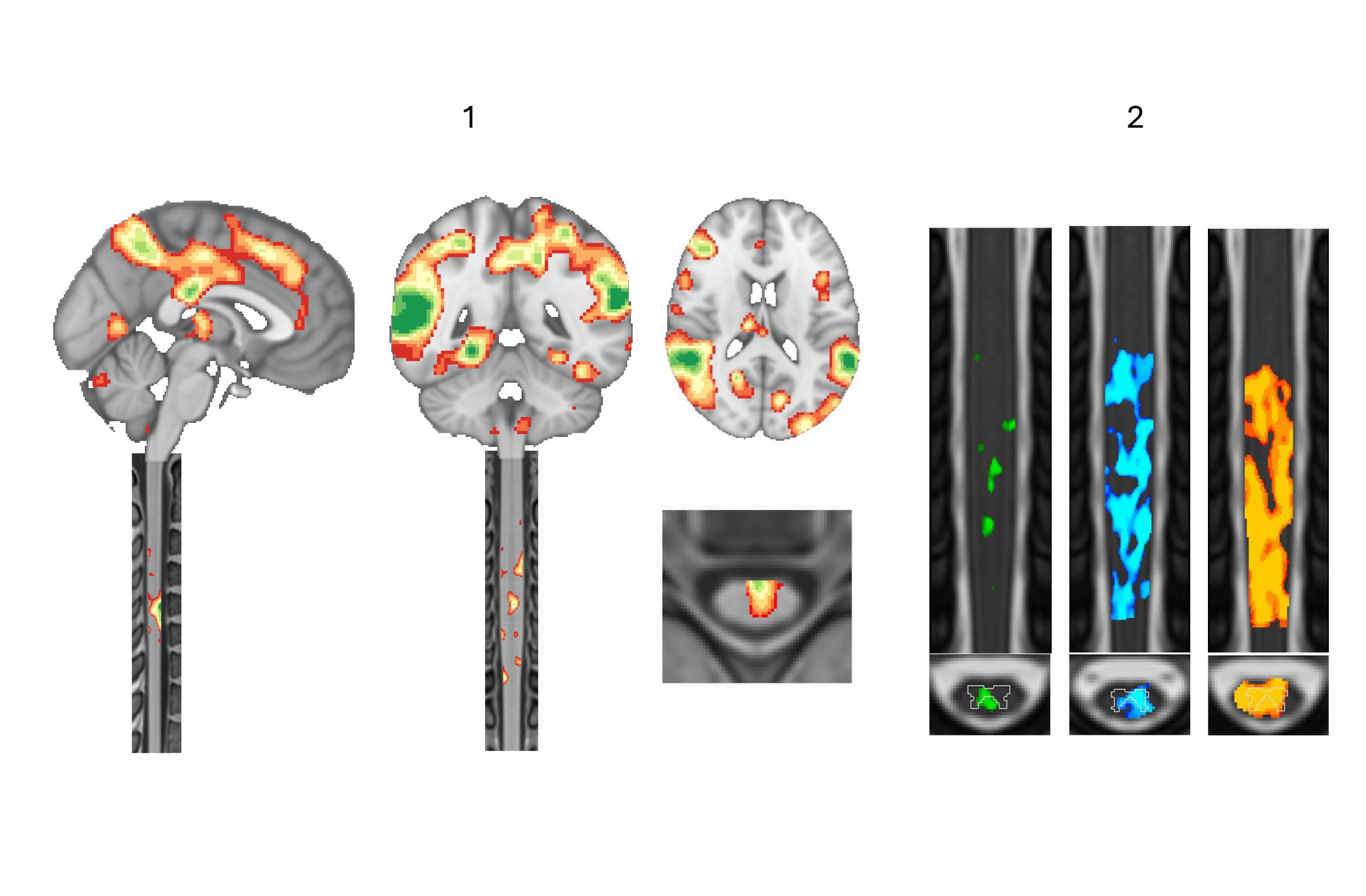
One recent study from Van De Ville's lab, now a preprint (a scientific manuscript without peer-review and that has not yet been accepted by a journal) takes that idea to the scanner. Working with Friedhelm Hummel's team, doctoral student Ekansh Sareen used transcranial magnetic stimulation (TMS), a coil held against the scalp that sends a brief magnetic pulse into the motor cortex, while imaging the spinal cords of 22 volunteers. It is the first time anyone has watched, in humans, what a pulse to the brain does in the cord. Weak pulses lit up only the cord's sensory side. Stronger ones spread through an intermediate zone to the motor neurons that make the hand twitch. "The spinal cord is not just an information highway," Sareen says. An information railway may be a closer analogy: the trains come to and from the brain, but some switches are thrown along the line that change the way information flows. "Beyond the cortex is sort of our core motto."
From lab to clinic
None of this is a therapy in itself. What imaging offers is a tool. Every spinal cord is wired a little differently, explains Sergio Daniel Hernandez-Charpak, imaging and computation manager at .NeuroRestore, the Lausanne research centre led by EPFL professor Grégoire Courtine and CHUV neurosurgeon Jocelyne Bloch. The nerves serving a given muscle can join the cord a segment higher or lower from one person to the next, and a tall person does not necessarily have a long lumbar spinal cord. For an implant, that matters. In 2022, .NeuroRestore reported that three people with complete paralysis could stand and walk again with a stimulator placed on the spinal cord. The .NeuroRestore team is now bringing the method to Lausanne's university hospital, CHUV, ahead of a trial in stroke, where patients, unlike many with spinal injuries, have no metal plates in the spine to blind the scanner. Hernandez-Charpak is candid about the limits: spinal fMRI, he says, is still "in its infancy."
Kinany and Van De Ville were among the authors of the 2022 landmark study: spinal fMRI, recorded while the participants' legs were stretched or their tendons vibrated, showed which segments answered to which muscles, and personalized models built on those maps guided where the electrodes should go. Surgery confirmed the predictions. Van De Ville sees further uses: spotting changes in the cord before symptoms appear, tracking a drug's effect over months, choosing which patients will benefit. "It will take sophisticated tools to select the right patient," he says.
Four hundred years after Galileo shook the world with his Moon drawings, we now have powerful telescopes and computers doing the signal processing. But scientific progress is not linear; it moves in leaps and jolts. Taking neuroscience beyond the cortex is one of these small leaps. And it started because someone saw that there was a missing piece to the puzzle. A piece that, if found, could one day help someone get back some of what they lost in an accident, a stroke or a neurodegenerative disease.
In the café where we sit, I spend an hour filling Kinany in on the different directions research on spinal cord imaging has taken since she left. "I put eight years of my life into this," she says. "So, it's nice to see that it's still moving forward."