Real-time monitoring of neuronal activities, intended for exploring the mysterious function of the brain and understanding the underlying mechanisms of neurological disorders such as epilepsy and Alzheimer's disease, has long posed a formidable challenge.
Potassium ion (K+), as a key determinant of membrane potential, has been one of the main investigation targets because its concentration change in extracellular space directly affects the membrane potential of the neurons and impacts the neuronal intrinsic excitability and synaptic transmission. Among a myriad of monitoring methods, non-invasive fluorescence detection of [K+]o holds the potential to convey the spatial and temporal information about [K+]o, manifesting itself as a promising candidate to unravel the complex neural interactions at multiple scales in the brain.
The currently available optical K+ sensors have so far allowed the routine measurements of spatiotemporal dynamics of [K+]o in cultured cells, mouse brain slices and anesthetized animals. Although imaging [K+]o dynamics in waking and freely moving animals is imperative for direct comparative analyses of their behaviors and neural activities, the existing optical K+ sensors fail to fulfill these functions due to two limitations. First, they are not sensitive enough to detect a small [K+]o change. Second, despite significant efforts to improve the selectivity of K+ reporters, they are still far from satisfactory in differentiating K+ from sodium ion (Na+).
On February 10, Prof. LING Daiyun from Zhejiang University, Prof. Taeghwan Hyeon from Seoul National University and Prof. CHEN Zhong from Zhejiang University, etc. co-published a research article entitled "A sensitive and specific nanosensor for monitoring extracellular potassium levels in the brain" in the journal of Nature Nanotechnology.
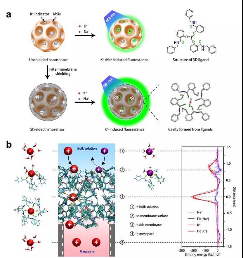
In this study, researchers develop a highly sensitive and selective K+ nanosensor that can monitor the changes of [K+]o in the brain of freely moving mice. They achieve the sensitive optical readout of [K+]o changes by incorporating commercially available K+ indicators into mesoporous silica nanoparticles and subsequently shielding the MSNs with a precisely controlled thin layer of three-dimensional tripodal ligands. The ligands constitute a K+-specific filter membrane that contains a binodal 3,6-connected network with a pyrite topology. The pores (~5.7 Å in diameter) of the network allow only K+ (~2.66 Å in diameter) to diffuse into and out of the mesopores.
This K+ nanosensor can monitor potassium changes in the brain of freely moving mice undergoing epileptic seizures. An optical potassium indicator is embedded in mesoporous silica nanoparticles shielded by an ultrathin layer of a potassium-permeable membrane, thus preventing the diffusion of other cations and allowing the specific capturing of potassium ions. The shielded nanosensor enables the spatial mapping of potassium ion release in the hippocampus of freely moving mice.
In contrast with the invasive K+-selective microelectrode, which can be used merely for the single-point measurement of [K+] in a fixed sample, this type of nanosensor is non-invasive and can convey the spatial information of [K+] change on a wide scale for freely moving mice. Further development of tissue-penetrating near-infrared emission-based K+nanosensors would allow the precise detection of the epileptic fociin whole-brain imaging, thereby facilitating diagnosis and therapy of epilepsy and decreasing the need for resection surgery in epilepsy. Once loaded with antiepileptic drugsand coated with nanocomposites that can be disrupted by elevated [K+]o, this type of nanosensor will also enable a highly localized and on-demand drug release at seizure foci.


