
Brown University engineers developed a microchip that tracks cell movement using electrical capacitance rather than optical lenses, which could aid in understanding processes such as cancer metastasis, wound healing and immune response.
PROVIDENCE, R.I. [Brown University] - Engineers from Brown University have developed a new way of imaging cell migration dynamics that are important in understanding critical processes like cancer metastasis, wound healing and immune response.
The technique, described in a study published in the journal Lab on a Chip, uses an array of one million microscale electrodes on which cells can be placed in a liquid nutrient solution. The system tracks the cells' movement through electrical capacitance, making the technique a promising alternative to more complicated optical microscopes and fluorescence imaging, which can damage cells and potentially disrupt their behavior.
"Moving cells are very challenging to track since they change shape and move rapidly, so computational analysis can be quite error-prone." said Hyuntae Jeong, a postdoctoral research associate in Brown's School of Engineering and co-first author. "Cells often have to be labeled with fluorescent dyes in order to see them, but dyes can cause DNA damage, which could affect the way they move or even cause cell death."
The new technique can track cells without disturbing them and without hours staring through a microscope. The microelectrodes are arranged on a square microchip with sides about a half inch in length. When cells placed on the chip grip the surface to move, the contact affects the electrical properties near the surface of the chip. By measuring tiny changes in electrical capacitance, the device can precisely track each cell as it moves across the array.
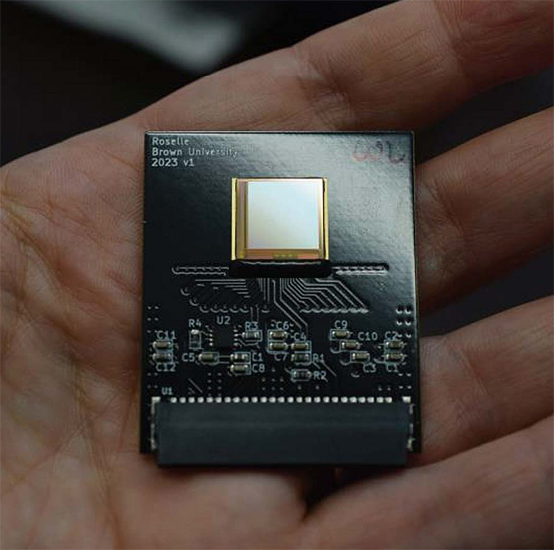
"By using capacitance rather than light, our sensor lets us observe cell migration without labeling, which may decrease unwanted artifacts," said co-first author Pushkaraj Joshi, a former postdoctoral research associate in engineering who is now a senior research associate in Brown's Department of Physics.
Jacob Rosenstein, an associate professor of engineering who co-led the study, says the approach is a bit like fingerprint scanners used to authenticate people's identity.
"There are differences in electrical capacitance between the ridges in a fingerprint and the spaces in between, and that's what some scanners read," he said. "It's a similar concept here. We're detecting the differences in electrical capacitance between where cells are touching the surface and where they are not, as they move over time."
In laboratory tests, the researchers showed that the technique could successfully track the motion of human breast cancer cells and capture cell division events as they occurred. The study also showed that the chip could reveal the dynamics of multicellular spheroids, capturing the motion of "leader cells" at the outer edges of clusters as those cells begin migrating outward. In addition, the researchers successfully imaged honeycomb tissues comprised of hundreds of thousands of cells to demonstrate that the technique could resolve voids and boundaries due to complex changes in shape.
The device could be mass produced fairly inexpensively, the researchers say, and it has several advantages over traditional microscope imaging techniques. The relatively large sensor area enables imaging of large tissues without the need to stitch together multiple images, which is often necessary with optical microscopy. And in addition to eliminating the need for fluorescent labeling that can damage cells, the system is also easily portable.


