Researchers from the School of Biomedical Engineering & Imaging Sciences have published a new study exploring the use of Positron Emission Particle Tracking (PEPT) in a living subject for the first time.
PEPT technology allows for the 3-D localisation and tracking of a single radioactive particle within large, dense, and/or optically opaque systems, which is difficult to study using other methodologies. The technology is currently used to study flows within complex mechanical systems such as large engines, industrial mixers, etc., but has not yet been translated for use in biomedical applications.
PEPT has previously been an unexplored area in biomedical imaging due to the lack of methods to isolate and radiolabel a single particle of a small enough size and with enough radioactivity which to would enable it to be injected and detected in a living subject.
In this new study published in the journal Nature Nanotechnology, main author Dr Juan Pellico and a multidisciplinary team led by Dr Rafael T. M. de Rosales were able to synthesize, radiolabel and isolate a single submicrometer particle of silica with sufficient radioactivity to allow detection with both standard PET imaging and PEPT for the first time.
Our ambition is to further develop these findings and developed improved PEPT tracers that will allow us to fully explore the potential of PEPT in biomedicine to provide whole-body information about blood flow dynamics in different settings, with unique applications such as the study of complex multiphase flow of blood, crucial in clinical physiology and drug delivery. Other potential applications include using single particles for high-precision PEPT-guided radiotherapy or surgery. Moreover, in vivo PEPT with single radiolabelled cells should allow the evaluation of the motion and migration of individual cells, and their interaction with blood vessels and tissues.
Dr Rafael T.M. de Rosales, Reader in Imaging Chemistry at the School of Biomedical Engineering & Imaging Sciences
"PEPT allows you to triangulate the position of the single particle inside the body with high precision and in real time," said Dr T. M. de Rosales.
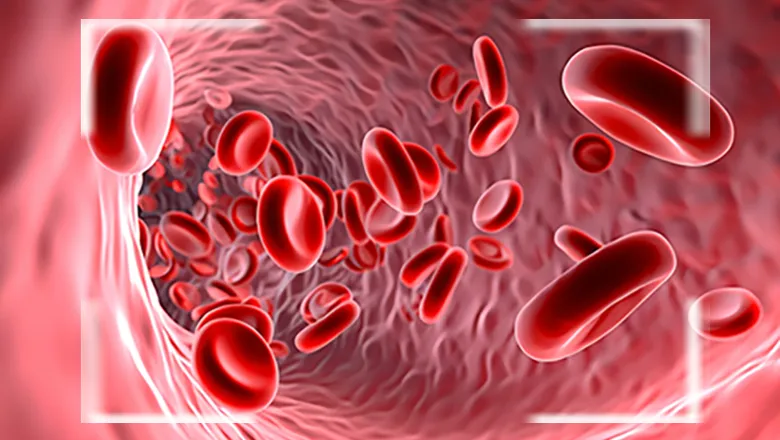
"In current PET imaging methods, we inject billions or even trillions of radiolabelled molecules into the patients and the resulting images represent their average distribution after a period of time, usually 10-30 minutes. This does not give you information about the velocity of these molecules or their exact location inside the body in real time, which could be useful for the study of haemodynamics, or how blood flows through your vessels.
"PEPT, by tracking single particles in real time, should allow the study of the velocity, density, and overall dynamics of blood flow that are currently impossible to study by any other imaging modality. The study of haemodynamics at the whole-body level is particularly timely since clinical total-body PET scanners are now available, one of which will soon be installed here at King's."
In vivo PEPT has the potential to provide important breakthroughs in the evaluation of abnormal events in cardiovascular diseases or cancer where the blood flow has a prominent impact. Future clinical applications may include the detailed analysis of blood flow and pressure gradients within lesions such as tumours or vascular lesions, where blood flow is abnormal, which could be used to guide treatment options for patients.
In this story

Reader in Imaging Chemistry