A state-of-the-art facility where researchers can explore biological processes and new materials at the molecular level opened recently on the first floor of the Frost Institute for Chemistry and Molecular Science at the University of Miami.
Named the Advanced Tools for Observing Molecules and Materials, or the ATOMM facility, the lab's five high-performance electron microscopes allow researchers to observe and explore biological and material systems at a minuscule scale. This is a key part of the development process for creating new therapeutics or novel materials that may help produce smaller, more energy-efficient batteries, computer chips, or even new substances that make airplanes and spacecraft safer and lighter.
"These are instruments that can detect and characterize what is happening at a very, very high resolution between atoms in materials and in biological systems, which can lead to new discoveries and new drugs that can be used in humans," said Sylvia Daunert, co-interim director of the Frost Institute for Chemistry and Molecular Science and chair of the Department of Biochemistry and Molecular Biology at the Miller School of Medicine. "You can see what happens with these materials, how they are built, and what kind of architecture they have. This is important because knowing how they are built structurally can lead to new materials that can be used in many different capacities."
The shared facility is open to University scientists but will also be available to private industry and other institutions looking to conduct electron microscopy research.
Video: Matthew Rembold/University of Miami
"We hope that University researchers, as well as other researchers in the community, will take advantage of these amazing new tools to advance scientific discovery at our world-class facility," said Marc Knecht, co-interim director of the Frost Institute and chair of the Department of Chemistry.
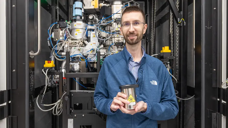
Electron microscopy is a cutting-edge imaging strategy used by instruments in the ATOMM facility that helps researchers understand the interactions between individual molecules using a focused beam of electrons to help visualize samples. The five specialized instruments at the Frost Institute were all produced by Thermo Fisher, and each is housed in its own individual room, with 10-foot ceilings built to accommodate the equipment. Many are geared toward biological research and drug discovery, but two of the instruments also provide strong insight into materials science work, said Maciej Jagielnicki, facility manager of the ATOMM facility.
The instruments are set up in order of their ability to capture finite details, Jagielnicki added.
"As you move up to each new microscope, you are able to get more detailed information on whatever you are studying," he said.
The first microscope, the Talos F200C, is an accessible and durable transmission electron microscope, well suited for routine imaging and screening of biological and materials science samples. It provides reliable, high-quality images with resolution appropriate for a broad range of research needs, facility managers noted. The second instrument, the Spectra 300, is the center's most advanced microscope for materials science research, offering state-of-the-art resolution, as well as multiple analytical capabilities for studying structure, composition, and chemistry at extremely small scales. The Krios G4 and Glacios 2 microscopes are specialized cryogenic electron microscopes, or cryo-EM instruments, designed primarily for biological and molecular samples that are rapidly frozen and imaged at extremely low temperatures. The final instrument, Aquilos 2, is not just a typical imaging microscope; it is a cryo-focused ion beam system that is used to prepare ultrathin frozen sections of samples, enabling their internal structures to be studied in greater detail.
In addition:
- Talos F200C is a screening microscope for soft and hard materials and gives researchers information about whether a project is feasible. For example, Jagielnicki said, scientists can see "if a potential drug molecule will bind to proteins in the body." It can also give good atomic resolution of a materials science sample, such as a metal-organic framework, for instance," added Muchuan Hua, a staff scientist and electron microscopy expert focused on materials science at the ATOMM facility. Before coming to the U, Hua worked at Argonne National Laboratory, where today's lithium-ion batteries were developed.
- Spectra 300 is a top-of-the-line electron microscope for materials science and can help researchers "distinguish individual atoms," said Knecht. "With this tool, you can literally pinpoint gold and silver atoms, where they are in the material, and see what they are doing. We need to know this information because where atoms are oriented can change the properties of a material, which can also change the reactivity, or how it catalyzes a chemical reaction," he added.
- Krios G4 is a microscope used for illustrating proteins in the body and their interactions with molecules, a key part of the testing process for therapeutic drug discovery.
- Glacios 2 offers insight and high-resolution images that reveal the structure of complex proteins and macromolecules.
- Aquilos 2, housed on the second floor, enables researchers to observe cellular structures from various angles.
There are many things that make the ATOMM facility unique, University scientists noted. What sets it apart from the University's other facilities at the Miller School of Medicine is that after biomedical researchers formulate a new therapeutic, they can then test it at the ATOMM facility with cells from the area of the body that it is designed to impact and see whether this interaction is beneficial, Daunert said. The facility is also distinct because it offers electron microscopy for both materials and biological samples, Jagielnicki and Hua added.
"These are some of the most powerful tools human beings have made for getting the finest resolution and images of anything you create, and they allow you to directly analyze how atoms are constructed in molecules," Hua said. "Since we want to make chips smaller, this could help us to examine how to make semiconductors even smaller, show where the defects are, and also how the conductivity is preserved to perfect its properties."
Iga Kucharska, research assistant professor of chemistry and a structural biologist, is helping to manage the ATOMM facility. But she is also working to understand how some of the body's most vital protein groups operate so she can develop drugs to combat the spread of heart disease and cancer. Kucharska is eager to utilize the ATOMM facility more for her research.
"Electron microscopy helps you understand how and where the drug molecules bind to proteins—knowing this can help you to develop even better more effective drugs because the process allows you to understand these interactions at the atomic level, so you can then design therapeutics that will bind even better," Kucharska said.


