In today's scientific world, microscopic images have become a powerful resource for research. With access to advanced microscopes, researchers can now create unique images of structures and objects. Beautiful and captivating images that can also convey complex context to a wider audience.
Microscopic images offer a clear advantage over purely quantitative measurements: they allow us to see the structure of an object in a way that numbers and diagrams cannot. By using advanced microscopes, scientists can create images that show details invisible to the naked eye. This makes it possible to explore and explain complex structures and phenomena that are also visually easy to understand.
New insights about the body
Medical research tends to become more and more complicated. This is according to Tomas Björklund, Principal Investigator at the strategic research area MultiPark, a translational research network on Alzheimer's disease and Parkinson's disease.
- This is largely because we also use very large amounts of data and advanced computer models and AI to analyse and interpret the data. Therefore, I would say that communication with images has never been as important as it is today. It's only when we see the results in our microscopes that I feel completely confident that all our computer models are right and that it really works in the real world.
Can this type of imagery provide new insights about the body that can drive research forward? This is a crucial question for many researchers in biology and medicine.
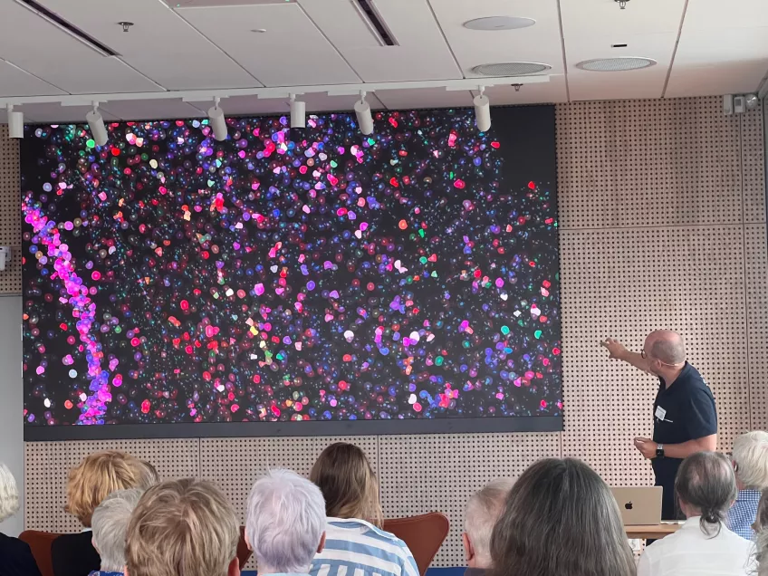
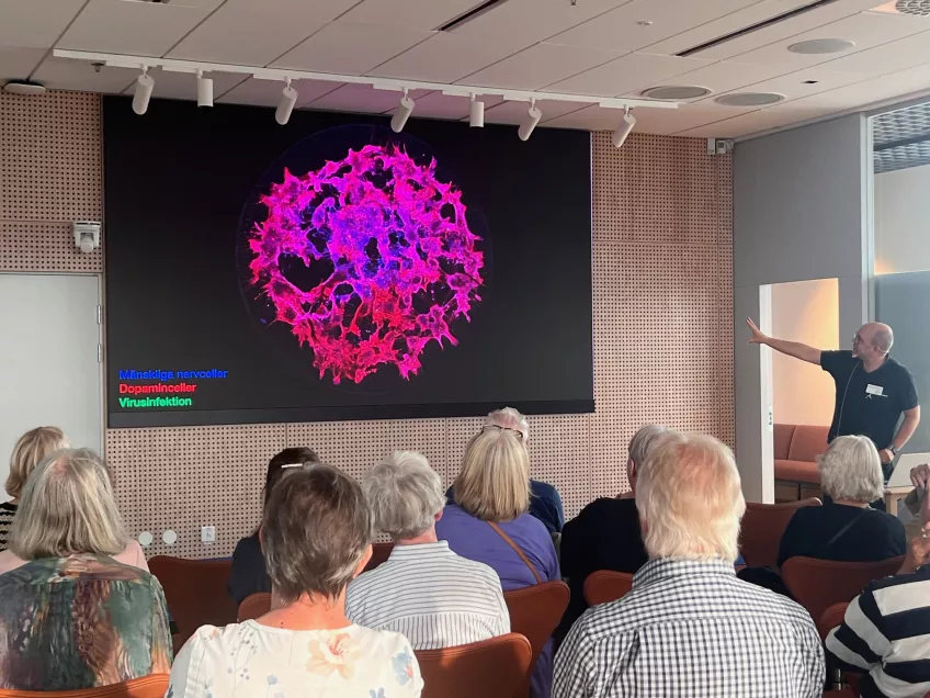
- It is still often the case that it is through the microscope that we make the big new discoveries or see that new treatments work. For example, we had been working for four years on developing new synthetic viruses to effectively infect dopamine cells for the treatment of Parkinson's disease. Up to this point, all development had been done in test tubes and in computer simulations and this was the first time we tested these new viruses on human neurons grown in a petri dish.
In one dish, the neurons were infected with the virus used so far in new treatments for Parkinson's disease and the researchers saw almost nothing.
- This was expected. But when we moved the microscope to the second dish infected with our synthetic virus, they dissolved with an almost blinding light. This was a magical feeling and is the kind of reward that gives us energy to keep working.
A similar feeling that Tomas Björklund thinks is difficult to produce when the analysis takes place without images, for example when analysing measurements on the computer, although these results can also be just as important.
The body's innermost room
Tomas Björklund also wants to reach out communicatively with images from the lab and was therefore involved in the project 'The invisible body' at Sven Harry's museum in Stockholm in 2017. A public exhibition that showed magnificent images from the innermost parts of the body.
- We were invited to submit a visually and scientifically appealing image. We chose to contribute an image that meant a lot to us and it became a really nice exhibition with many beautiful images.
The Ragnar Söderberg Foundation, which was behind the exhibition, donated the enlargements to the respective universities and now Lund University's contribution hangs together with several other images from the lab in the public spaces at the Biomedical Centre (BMC) in Lund.
- The response from the exhibition at Sven Harry's museum was positive, but there was a limited number of people who could see the exhibition because it was in Stockholm. I'm glad that people can see these images here in Lund. Since then, we have received more positive reactions.
Qualitative information
Ellinor Molnár, a former researcher at Multipark, is also interested in the fascinating world of advanced microscopes that open doors to a new era of understanding and communication. Using microscopes, Ellinor Molnár and her research colleagues were able to identify more structural details such as specific location, shapes, colours and patterns.
- Firstly, microscopic investigations can provide more qualitative information about the structure of objects than purely quantitative measurements can. Secondly, it can be easier to understand what a microscopic image shows than what a mass of data means, especially for a person less familiar with the subject. This allows you to reach more people with your research and attract the interest of more than just experts in the specific field. It can also be beneficial from a teaching point of view, says Ellinor Molnár.
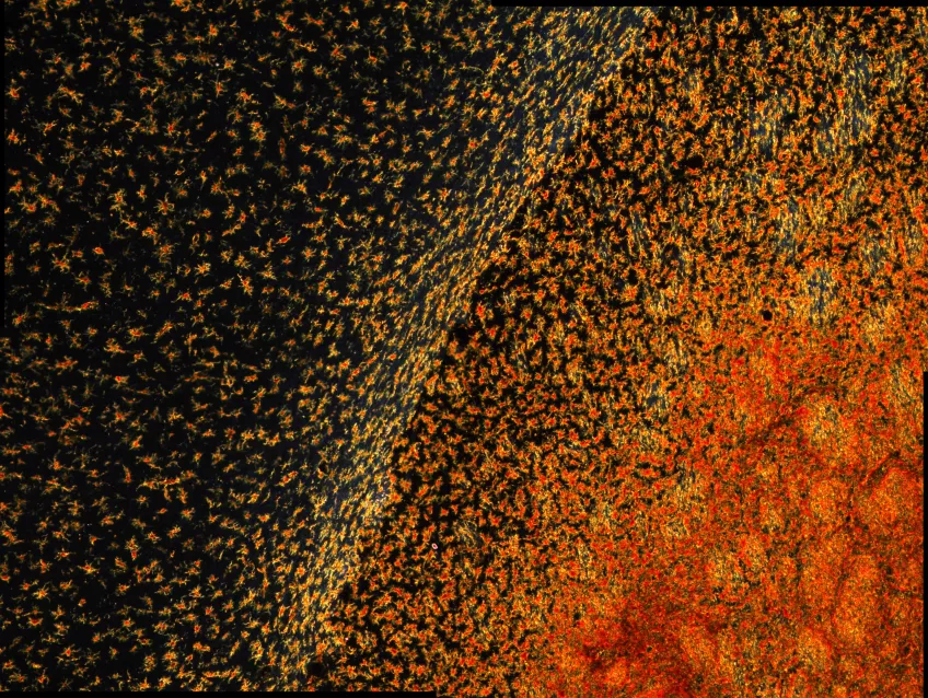
She entered the Multipark photo competition and won with a picture showing part of a rat brain that has been transplanted with human embryonic stem cells to treat Parkinson's disease.
What did it give you?
- I'm happy that I participated and proud that I won - I didn't expect that! I got super positive feedback from my lab colleagues and family members, which was great. It gave me an extra boost of confidence.