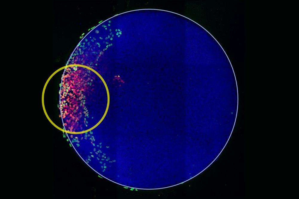
Scientists in the Brivanlou lab used light-inducible gene expression (yellow circle) and embryo models to demonstrate that, at the start of gastrulation, body-axis formation requires an interplay between biochemical signals and mechanical forces. (Credit: Brivanlou lab)
Only two weeks after fertilization, the first sign of the formation of the 3 axes of the human body (head/tail, ventral/dorsal, and right/left) begins to appear. At this stage, known as gastrulation, a flat and featureless sheet of cells folds into a living blueprint for the body, a fleeting transformation into axes and layers that will determine how every tissue develops. This all-important moment has, however, long stood beyond the reach of science, occurring too early and deeply within the uterus to study directly.
Now, a new study reveals that this pivotal step in human development is guided by a precise interplay between chemical signals and physical forces. Published in Cell Stem Cell, the paper introduces a light-based synthetic embryo tool that allows researchers to activate key developmental proteins known to initiate gastrulation. When the team used light to trigger one of these proteins, BMP4, they found that chemical cues alone were not enough-the transformation began only when the cells were also under the correct mechanical conditions. The results reveal a fundamental interdependence between tissue mechanics and molecular signaling, offering a more faithful model of early human development and a potential foundation for future regenerative and fertility therapies.
"We can now generate self-organization and different cell types, just by shining light on it," says Ali H. Brivanlou, head of the Laboratory of Synthetic Embryology. "This allowed us to make a major discovery about the role of mechanical forces in embryonic development."
Optogenetics advance sheds new light
Gastrulation begins with symmetry breaking. A uniform sheet of embryonic cells organizes into a three-dimensional head-to-tail axis-the spatial blueprint that determines where the head, spine, and limbs will eventually form. Brivanlou and colleagues have been chipping away at the mystery of this key developmental stage for decades, with the help of animal models and laboratory studies of human embryonic stem cells. "Gastrulation happens in the uterus shortly after implantation, so it cannot be studied without the use of human pluripotent stem cells, in vitro," says Riccardo De Santis, director of the Human Pluripotent Stem Cell Resource Center at Rockefeller and co-first author of this study, together with theoretical physicist Laurent Jutras-Dubé. "Our goal was to open a window into a moment of development that cannot otherwise be studied in vivo."
Prior work demonstrated that biochemical signaling molecules, such as BMP4, influence the behaviors of cells and tissues to regulate embryonic development. But studies in frog and chick embryos suggested that this was only part of the story. Mechanical tension, tissue geometry, and various physical forces also appeared to play a role in ushering animal embryos through development. "A lot of data is finally coming in, and it's now clear that the role of mechanical signaling has been underappreciated," De Santis says.
De Santis developed an optogenetic tool that allows the team to investigate the interplay between biochemical signals and mechanical forces, within the context of human development. By engineering human embryonic stem cells to respond to light, his system allowed researchers to activate developmental genes with extraordinary precision. When exposed to a specific wavelength of light, the cells were designed to flip a genetic switch that permanently turns on BMP4. This setup also allowed scientists to choose exactly when and where the signal is activated in the clump of embryonic cells, enabling them to test, for the first time, how tissue geometry and mechanical stress at any physical location in the embryo could influence development.
The rise of mechanical forces
When the team used this light-based system to activate BMP4 signaling in human stem cells, the role of mechanical forces quickly became clear. In cultures where BMP4 was triggered in unconfined, low-tension environments, gastrulation never fully coalesced. BMP4 alone was enough to give rise to extra-embryonic cell types, like those that form the amnion, but the sample failed to generate the mesoderm and endoderm, the layers that go on to build the body's organs. This demonstrated that morphogens alone are not enough to accomplish gastrulation.
But when the team pointed their "remote control" at the edges of confined cell colonies, and to cells embedded in tension-inducing hydrogels, gastrulation's missing layers began to form. Further experiments revealed how mechanical tension via YAP1 fine tune the downstream biochemical signaling pathways mediated by WNT and Nodal, which tell cells what types of tissues to become. A previous study led by Senior Research Associate Francesco Piccolo, in collaboration with the late Jim Hudspeth, head of Rockefeller's Laboratory of Sensory Neuroscience, demonstrated that the nuclear levels of the mechanosensory protein YAP1 play a crucial role in regulating self-organization in micropatterns (Piccolo et al., 2022). The present study unveiled that nuclear YAP1 acts as a molecular brake of gastrulation, preventing these transformations from occurring too soon. The results suggest that gastrulation can begin only when molecular signals and mechanical tension align-cells, it seems, must be both chemically prepared and physically primed.
"There has been so much beautiful molecular biology on the embryo, so much incredible work on signalling. But we have, as a field, neglected physical forces," Brivanlou says. "It is now clear that, without mechanical forces, we cannot generate cells for proper embryonic development."
The results not only demonstrate the power of optogenetic tools and the importance of mechanical forces, but also provide a new framework for understanding how human embryos organize themselves at the very earliest stages. To complement the experiments, Laurent Jutras-Dubé developed a mathematical model that acts as a "digital twin" of a developing embryo. This computer simulation shows how biochemical signals like BMP4, WNT, and NODAL move through tissues and interact with physical forces. By using actual measurements of mechanical tension, the model can predict how signaling patterns and tissue organization lead to specific cell layers. The simulations closely match what was observed experimentally, demonstrating that both biochemical signals and mechanical tension must work together for this embryological signaling cascade to self-organize. This integrated approach provides a quantitative way to understand how the embryo changes during early development. Built on a microchip platform, these upgraded synthetic embryos build on landmark work from the Brivanlou lab which, in 2014, was the first to show that human embryonic stem cells grown on microchips could self-organize into two-dimensional "gastruloids" that mimic early developmental patterning.
Next, the team plans to explore the possible existence of a mechanical organizer-a force-based counterpart to classical signaling centers that shape the early embryo. They suspect that, in addition to chemical cues, the embryo must satisfy specific physical conditions to progress through developmental milestones-a state that the authors call mechanical competence. "The existence of a mechanical organizer is a provocative concept that could prove transformative," De Santis says.
Beyond its conceptual impact, the optogenetic remote-control embryo offers an unparalleled platform for experimentation, enabling light-driven control of developmental cues in engineered microenvironments. Such systems could advance regenerative medicine and reproductive health, from refining stem-cell therapies that activate on demand to illuminating why early pregnancies sometimes fail. "Our work focuses on fundamental biology and basic science, but the implications are really important in terms of supporting fertility," De Santis says. "When we improve our understanding of the underlying rules of embryogenesis, we can use that information to give people the best opportunities for building future families."
Already, the present work offers an unprecedented view of where we all began. "Sometimes scientists get lost in the tools and the chips and the lights, and we forget that this kind of research is special," Brivanlou says. "When I look at gastrulation, I feel like I'm looking at a mirror that reflects my own past. It's more than just science. It's an opportunity to look at where we all came from-that magical stage of development that makes us what we are."