Conventional MRI scans, familiar to us from hospitals, have a resolution of around one-tenth of a millimeter, which allows them to image incredibly thin slices of our bodies, from head to toe, helping physicians diagnose a variety of medical conditions. Even this ultra-high resolution, however, is insufficient for researchers who want to study the structure of individual molecules. New technology developed in Dr. Amit Finkler 's laboratory at the Weizmann Institute of Science allows researchers to conduct MRI scans at a resolution of one nanometer (one millionth of a millimeter, or one-billionth of a meter) or higher. The new nano-MRI device, described recently in Communications Physics , may make it possible to distinguish all the tiny particles that make up a single molecule, generating the most detailed images of individual molecules ever produced. It represents a massive leap forward in the race to develop nano-MRI applications for research and for use in the materials and pharmaceutical industries.

MRI is based on a property of elementary particles within atoms called spin. It's a magnetic property that can be visualized as a rotation around an axis, similar to a spinning top, and it is characterized by a frequency - the number of "rotations" per second - known as the resonance frequency. This resonance frequency is what the MRI device measures. It depends on the type of particle being measured and the strength of the surrounding magnetic field. In conventional MRI, a gradient magnetic field, which varies in strength along the patient's body, causes the resonance frequency to vary as well, allowing the machine to differentiate between slices of tissue. A steeper gradient allows for thinner slices. The question that the Weizmann researchers posed was: Could a gradient magnetic field also be used to distinguish the individual particles within a single molecule?
""The nano-MRI we are proposing can operate at room temperature and examine the structure of materials under the conditions at which they are supposed to be used"
Scientists in Finkler's laboratory in the Chemical and Biological Physics Department had already developed a method of magnetic resonance scanning based on a synthetic diamond. Inside the diamond is a tiny defect the size of a single atom, called the nitrogen-vacancy center. This defect acts as a sensor that changes the intensity of the red light it emits, depending on the spin of the adjacent particles. The advantage of the nitrogen-vacancy center is that it is highly sensitive to even the weakest signals - such as, for example, the presence of a single particle located 50 nanometers away. The problem, however, was that it didn't always differentiate effectively between adjacent particles, and the light it emitted was influenced by the average properties of all nearby particles. This meant that it was difficult to use this sensor to image the individual atoms that make up a molecule.
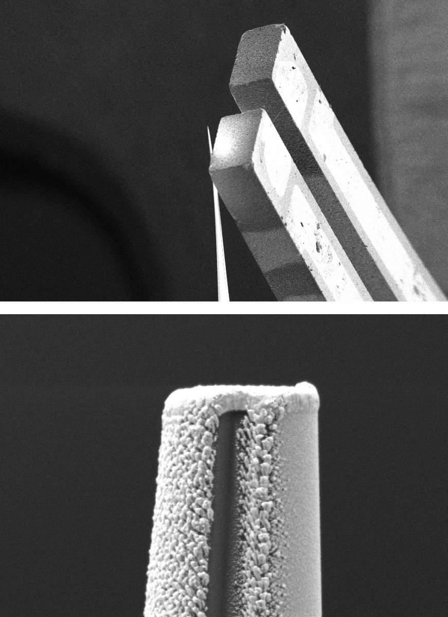
In their new study, led by doctoral candidate Leora Schein-Lubomirsky, the researchers developed a machine that generates a gradient magnetic field focused around an atomic magnetic sensor. This machine is based on a quartz tip coated with a gold conductor in the shape of a square arch. When an electric current was run through the wire, it generated a gradient magnetic field; the changes in the magnetic field were strongest near the corners of the rectangle and gradually weakened further away.
"The changes in the magnetic field led to changes in the resonance frequency of atoms, depending on their position in the molecule," Schein-Lubomirsky explains. "Previously, the sensor was incapable of distinguishing between several nearby hydrogen atoms and determining their locations, but now, within each region, a hydrogen particle will exhibit a different resonance frequency. We will then be able to assemble images showing these different locations into a complete picture of the molecule."
"Don't look at the field, but at the change within it"
"The realization that led to the new development was that we could produce a very strong gradient of the magnetic field even if its absolute size remained small," Finkler explains. "Even though our magnetic field is significantly smaller than that of a commercial MRI machine, its gradient - the rate at which the magnetic field changes with distance from the device - is much greater. This is how we obtained a resolution of one nanometer, and we believe our device is capable of reaching even higher resolutions - meaning it would be capable of scanning the structure of an individual molecule."
The new device is also an improvement over previous diamond-sensor systems, since it can activate and deactivate the magnetic field on demand - and does so within just 0.6 millionths of a second. This is because the field is not produced by a magnet, but rather by means of an electric current that can be turned on or off. "The ability to quickly activate and deactivate the magnetic field ensures that there are fewer disruptions and that the scan is more accurate," Finkler adds.
High-resolution molecular imaging plays a crucial role in the materials and pharmaceutical industries. Today, every manufactured drug undergoes magnetic resonance testing to ensure that it contains only the desired substance in the correct molecular structure and arrangement that is safe for human use. Current methods, however, require large sample quantities, which can be difficult to obtain, especially during early development stages. Additionally, these tests cannot be conducted at room temperature, and their resolution remains limited.
"The nano-MRI machine we are proposing can operate at room temperature and examine the structure of materials under exactly those conditions at which they are supposed to be used," Finkler emphasizes. "The machine will also produce a more detailed image of the molecular structure and make it possible to test a small and significantly cheaper sample containing only a few molecules of the material. Moreover, this device could help reveal why substances sometimes behave unexpectedly in the real world, compared to the results of laboratory testing, and whether there are unknown differences between substances that appear to be identical."

Science Numbers
The gradient of the magnetic field - the rate at which the field changes with distance from the device - is 0.1 tesla per meter in conventional MRI machines. In contrast, the newly developed device achieves a gradient of 1,000 tesla per meter, that is, a 10,000-fold increase that enables dramatically higher resolution.
Also participating in the study were Dr. Yarden Mazor from Tel Aviv University and Dr. Rainer Stöhr and Dr. Andrej Denisenko from the University of Stuttgart, Germany.
Dr. Amit Finkler is the incumbent of the Elaine Blond Career Development Chair in Perpetuity


