The Oplegnathus punctatus (O. punctatus) is an economically significant marine aquaculture species. It possesses a distinct beak-like tooth phenotype, allowing it to feed on hard-shelled foods such as oysters and sea urchins.
The jaw teeth of O. punctatus are fused with the upper and lower jaw bones, forming a robust parrot-like beak-shaped dental structure, with gaps between teeth filled with calcified material. Their beak-like healing teeth are a special type of jaw teeth that can regrow after wear and tear.
How are these unique beak-like teeth arranged and fused? What are the regulatory mechanisms behind their development? Now, a research team led by Prof. LI Jun from the Institute of Oceanology of the Chinese Academy of Sciences (IOCAS) reported their latest findings on the developmental pattern and continuous replacement regulation mechanism of the beak-like teeth in O. punctatus.
The study was published in International Journal of Biological Macromolecules on Aug. 9.
The researchers systematically investigated the developmental characteristics of the healing-type beak-like teeth in O. punctatus. For the first time, a "nested" arrangement pattern of healing teeth in O. punctatus was discovered, with a dental formula (4, 15-16, 10-1). The key developmental time points of the beak-like teeth were determined (28 dph, initiation of primary tooth germination; 40 dph, appearance of replacement teeth and start of dental fusion; 45 dph, commencement of ossification; 50 dph, completion of healing).
Furthermore, a total of 11 key genes (bmp2, bmpr2, smad1, wnt5a, msx, axin2, fgfr1a, fgfr2, pitx2, ptch1, cyp27a1) closely associated with the development of the beak-like teeth were identified, along with crucial regulatory pathways (Wnt, BMP, FGF, SHH). These genes and pathways collaboratively regulate the interaction between dental epithelium and mesenchyme during the pre-fusion, fusion, and post-fusion stages of dental fusion, promoting sustained proliferation and differentiation of enamel-forming cells and odontoblasts.
The researchers found that the cyp27a1 gene, closely associated with vitamin D metabolism and calcium accumulation, is localized in the upper jawbone and base of the beak-like teeth of O. punctatus. "During the rapid healing phase of the teeth, it accelerates its expression, promoting vitamin D metabolism, thereby regulating the differentiation of cells within the dental bud, as well as the mineralization of enamel and dentin in teeth of O. punctatus. This provides a calcium foundation for the healing and development of beak-like teeth," said MA Yuting, first author of the study.
"The study unveils the regulatory mechanisms of beak-like healing teeth development," said Dr. XIAO Yongshuang, corresponding author of the study. "It provides a new model for deeper understanding of fish dental development and adaptive evolution."
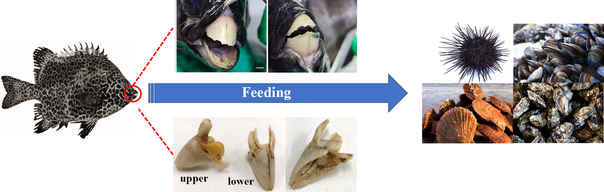
Fig. 1 External morphology of O. punctatus' beak-like tooth and feeding habits. (Image by IOCAS)

Fig. 2 3D reconstruction of beak-like healing tooth in O. punctatus based on micro-CT scanning (Image by IOCAS)
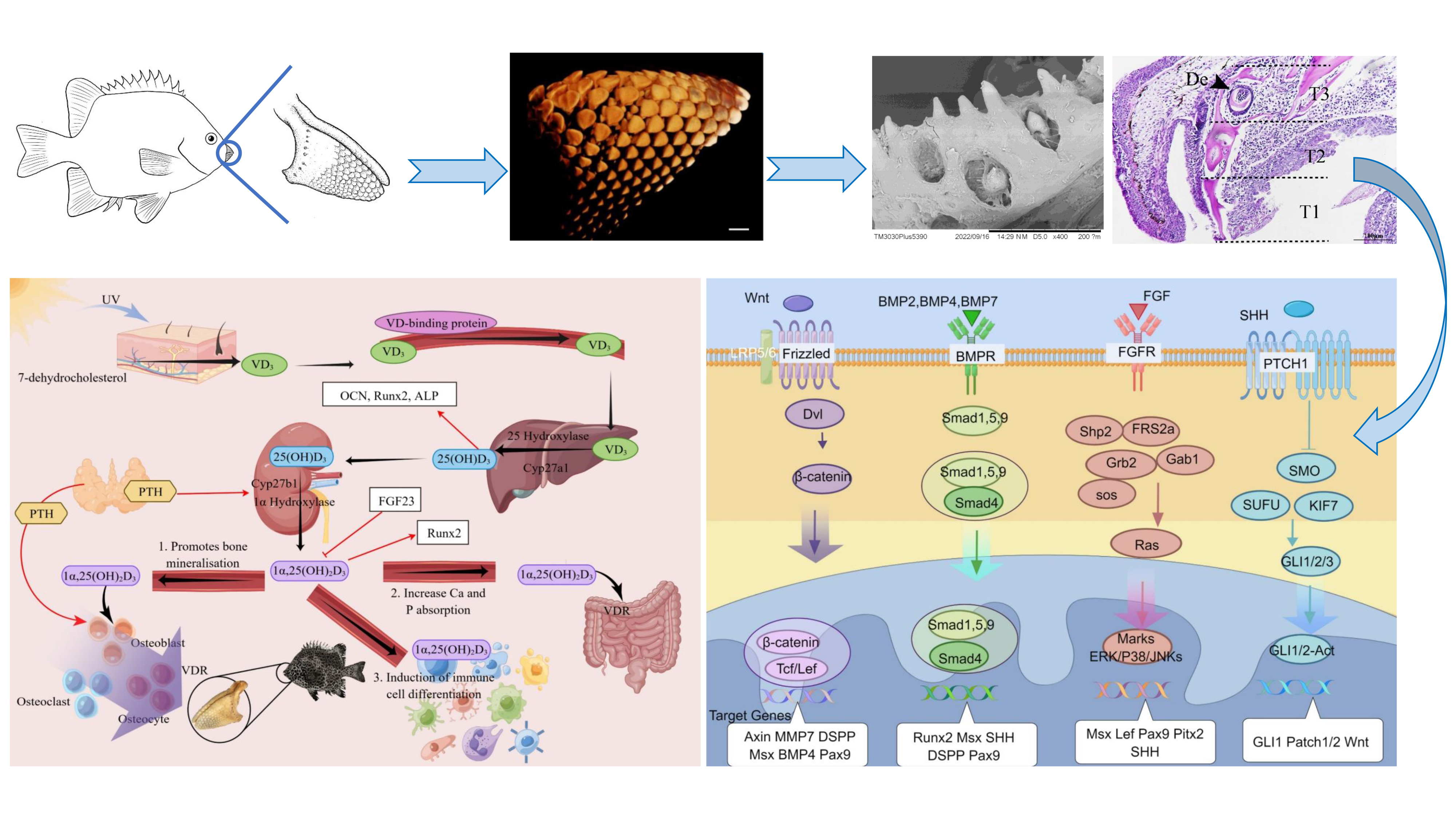
Fig. 3 Regulatory mechanism of development and formation of healing-type tooth in O. punctatus. (Image by IOCAS)


