Photoacoustic Imaging Enhances Ovarian Cancer Diagnosis Accuracy
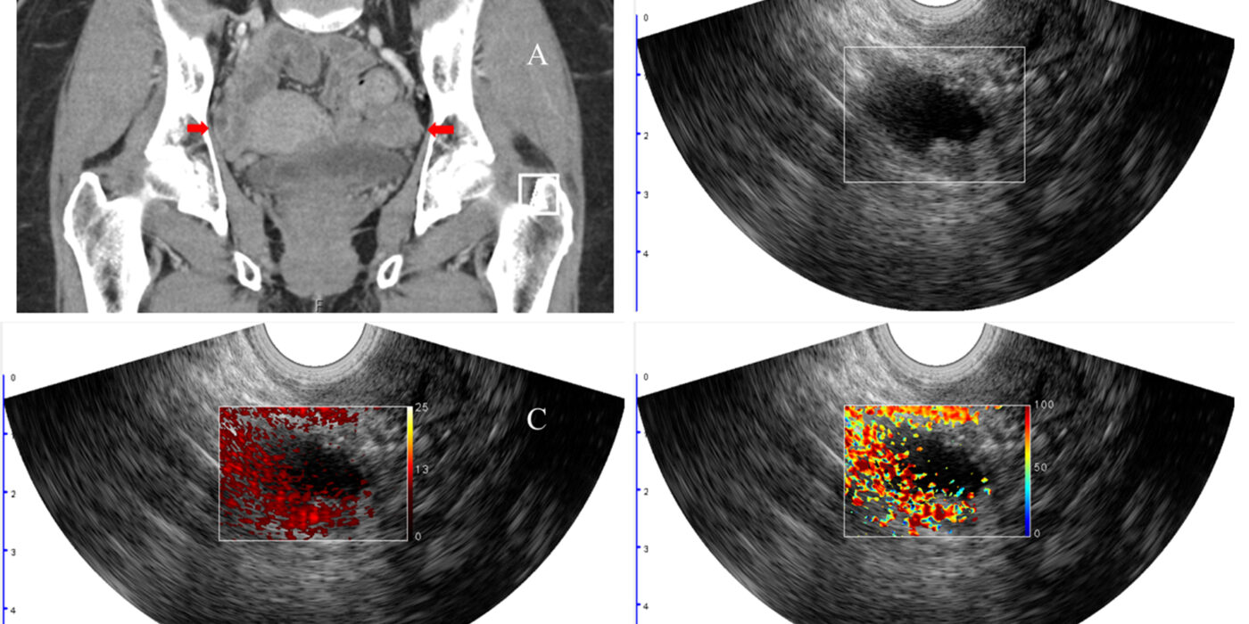
Quing Zhu, working with Matthew Powell, MD, and Cary Siegel, MD, from the School of Medicine, developed a new imaging method to better diagnose lesions in the ovaries and adjacent adnexa that may help to avoid unnecessary surgeries. This image shows a woman with pelvic mass and BRCA1 mutation. The arrows point to the right and left adnexa. Co-registered ultrasound shows a cyst with solid component in the right adnexa (B). Image C shows the relative total hemoglobin in the lesion, and image D shows the oxygen saturation, both of which are important predictors of malignancy. Pathology revealed a stage I high-grade serous carcinoma of the right ovary and fallopian tube. (Image: Zhu lab)
/Public Release. This material from the originating organization/author(s) might be of the point-in-time nature, and edited for clarity, style and length. Mirage.News does not take institutional positions or sides, and all views, positions, and conclusions expressed herein are solely those of the author(s).View in full here.