Developed at The Institute, the 3D heart-on-a-chip model reproduces key features of dilated cardiomyopathy and could help researchers identify more personalized treatments for cardiovascular disease.
Researchers at the Research Institute of the McGill University Health Centre (The Institute) have created a three-dimensional, beating heart-on-a-chip, or "mini-heart," using blood cells from a patient with dilated cardiomyopathy (DCM), a leading cause of heart transplantation. The new model could help researchers better understand the mechanisms underlying DCM, accelerate the evaluation of new treatments and, ultimately, explore the potential of patient-specific models to evaluate how an individual's heart tissue responds to different therapies. The study was recently published in Advanced Healthcare Materials.

Dilated cardiomyopathy is a serious heart condition in which the heart becomes enlarged and weakened, impairing its ability to pump blood and potentially leading to heart failure and abnormal heart rhythms. According to the authors of the study, it is associated with a 50 per cent risk of progression to heart failure within five years.
"With this new patient-derived 3D heart-on-a-chip model, we can begin to recreate aspects of an individual patient's heart disease outside the body, opening the door to applications in personalized medicine," says Ali Mousavi, first author of the study and a postdoctoral fellow in Dr. Renzo Cecere's Myocardial Regeneration Lab at The Institute.
Research in this field remains challenging, in part because conventional laboratory models, such as animal models and cells grown in the lab, cannot fully replicate the complexity of the human heart and cardiovascular disease, despite their essential role in research.

"This new model more accurately mimics the behaviour of a human heart than traditional research models do. This advance could help us find more effective treatments for dilated cardiomyopathy, ultimately reducing the need for heart transplants," says Dr. Cecere, Scientist in the Cardiovascular Health Across the Lifespan Program at The Institute, cardiac surgeon at the McGill University Health Centre (MUHC) and Associate Professor of Surgery at McGill University.
Building a patient's "mini-heart" in the lab
Heart-on-a-chip technology has advanced considerably over the past decade. The model developed at The Institute stands out because it uses cells derived from the blood of a patient with DCM.
"The blood cells are reprogrammed into induced pluripotent stem cells, which have the ability to develop into many different cell types in the human body. We then turn these patient-specific stem cells into heart muscle cells, or cardiomyocytes, and use them to create three-dimensional cardiac tissue on a chip. The result is a 'mini-heart' that beats and contracts on its own," explains Mousavi.
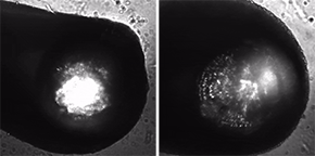
Another innovative feature of the study is the incorporation of tiny fluorescent beads directly into the tissues. These allow researchers to observe how different regions of the tissue move and contract with each beat, providing a more detailed picture of how the engineered heart tissue functions.
Recreating key features of heart disease
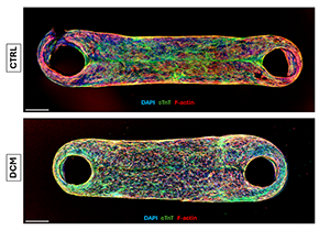
To evaluate the DCM patient model, the researchers created a second model using cells from a healthy participant and compared the characteristics and function of the two.
They found measurable differences between the two models in their structure, contractions, beating patterns, calcium activity and molecular characteristics. Notably, the tissues derived from the patient with DCM displayed abnormal, irregular beating patterns consistent with important features of the disease.
The researchers also exposed the tissues to norepinephrine, a substance that increases the heart's rate and force of contraction, confirming that the engineered tissues could respond to chemical stimulation.
"Together, these findings show that our model can reproduce important features of dilated cardiomyopathy and could eventually provide patient-specific insights into heart disease," says Mousavi.
In the future, the platform could also serve as a tool for preclinical drug development, helping researchers identify promising therapies earlier. Such models could also complement existing research methods and potentially reduce reliance on animal models for certain types of studies.
"This study provides an important proof of concept, and the next step is to expand the platform to a much larger number of patients," says Dr. Cecere. "Incorporating samples representing different genetic backgrounds and forms of DCM will be essential to understanding how the disease varies from one patient to another and moving this technology toward precision medicine."
About the study
The study Patient-Derived 3D Heart-On-a-Chip Model of Dilated Cardiomyopathy With Embedded Bead-Based Mapping of Tissue Contractilityby Ali Mousavi, Ludovic Mouttet, Shihao Cui, Yasaman Hekmatnia, Mehran Mottahedi, Ida Derish, Naimeh Rafatian, Mark Aurousseau, Gregor Andelfinger, Renzo Cecere and Houman Savoji was recently published in Advanced Healthcare Materials.
DOI: 10.1002/adhm.71464
The study was made possible thanks to the Courtois Cardiovascular Signature Program, supported by the MUHC Foundation, and its Heart-in-a-Dish project, a multidisciplinary research initiative aimed at advancing the understanding of DCM and the underlying causes of heart failure in individual patients.
The study was conducted as part of Ali Mousavi's visiting research in Dr. Cecere's laboratory at The Institute during his doctoral studies at Université de Montréal. It builds on Dr. Cecere's longstanding collaboration with Prof. Houman Savoji and his team at Université de Montréal in the development of biomedical innovations. Ali Mousavi is now a postdoctoral fellow at The Institute, supported by the Fonds de recherche du Québec (FRQ).