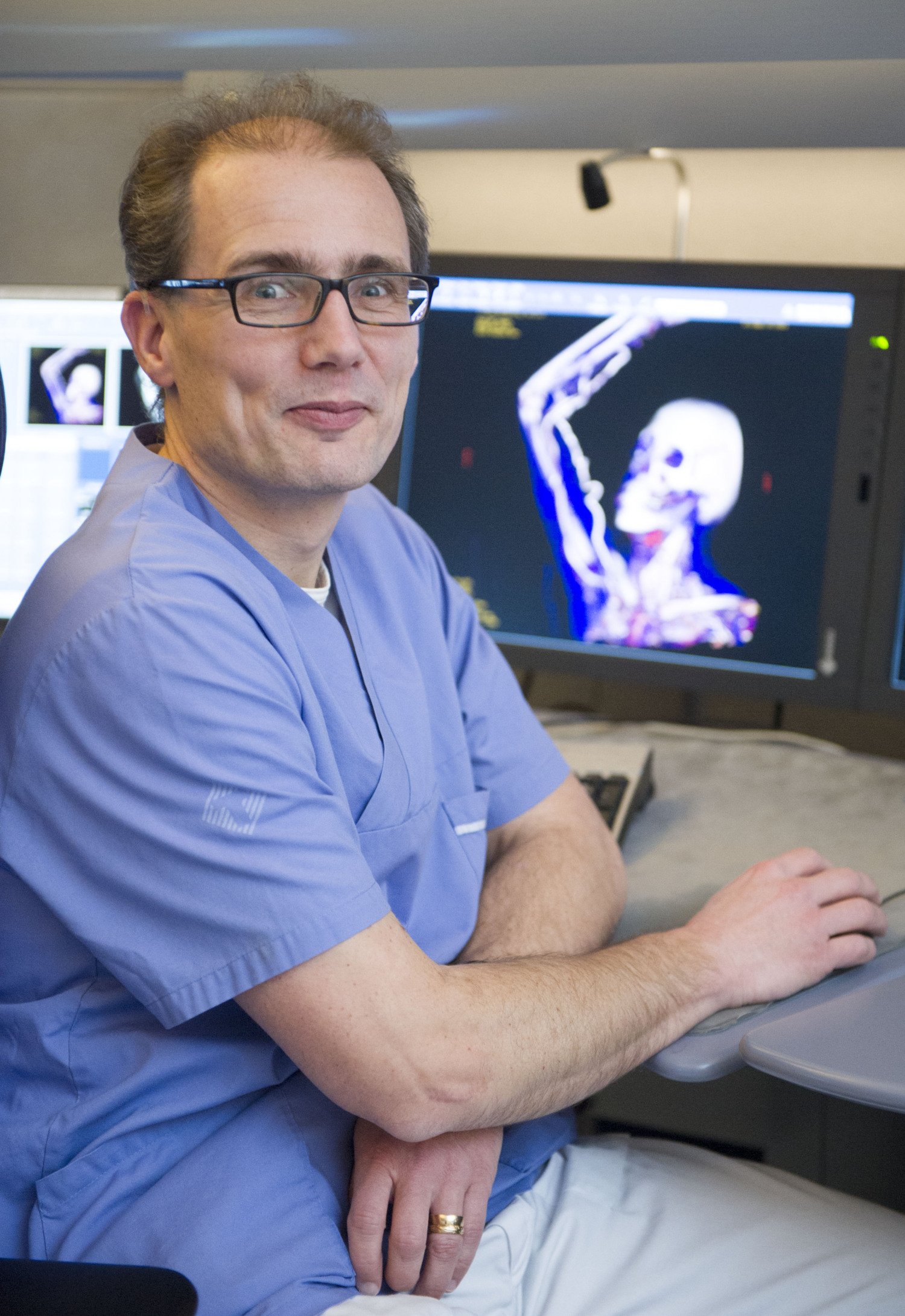
Can osteoporosis be detected in images taken for purposes other than fractures? According to radiologist Torkel Brismar, the answer is yes. He has explored this question in several studies.

Text: Annika Lund. Previously published in Medicinsk Vetenskap no. 3 2025 / Spotlight on osteoporosis
Osteoporosis is a silent disease that initially neither shows nor causes symptoms. Torkel Brismar , professor at the Department of Clinical Science, Intervention and Technology - CLINTEC, researches how screening for fragile bones could be implemented to identify patients before their first fracture.
'It is among the younger age groups, those between 55 and 70, that we have the best chance of preventing future spinal and hip fractures. Early treatment offers significant benefits,' says Torkel Brismar, who also works clinically as a radiologist.
He has investigated how hand X-rays can be used in this context. In an initial study, iust over 8,000 patients who had their hand X-rays between 2000 and 2008 were included. These patients either sought emergency care for suspected fractures or had been referred by rheumatologists.
Researchers assessed bone density in specific bones of the hand. They then tracked the patients over time using health registers and identified those who later sustained a hip fracture.
This provided insight into how a hand X-ray can help predict who is at of future fractures. In the next phase, researchers invited women to have their hands imaged during their mammografy-appointments. After the breast examination, the women were asked to place their hand on the plate of the mammography machine for an additional image, a process that only takes a few seconds. A total of 14,000 women agreed to participate. They also completed a questionnaire covering risk factors for osteoporosis, such as recent falls and mobility.
Predicting risk
The study revealed a clear link between bone density in the hand and fractures related to osteoporosis. However, the number of hip fractures was low, which is explained by the relatively young age of the participants (average age 53) and the short follow-up period (just over three years on average). "Still, the results are promising," says Torkel Brismar.
"Based on certain values in the bones of the hand, we can predict the risk of hip fracture within a specific timeframe, calculated at the individual level. It seems feasible to screen for osteoporosis in connection with mammography or when the hand is X-rayed for other reasons, such as after a fall," he says.
Researchers are now conducting a long-term follow-up to further understand the value of bone density in the hand. The women who had their hands X-rayed nearly two decades ago will be followed up again via health registers. The study will hopefully provide answers to how often the measurement should be repeated to identify as many cases of osteoporotic as possible.
In another project, researchers are training an AI to detect osteoporosis in CT scans taken during abdominal examinations.
"This opens up the possibility of making a more comprehensive assessment of patients' fracture risk. This is not assessed solely based on bone density - muscle mass and body fat also affect the risk of falling and breaking bones. Some patients may benefit from treatment other than medication, which primarily targets bone strength. This could involve training specific muscles, using hip protection trousers or other interventions", says Torkel Brismar.


