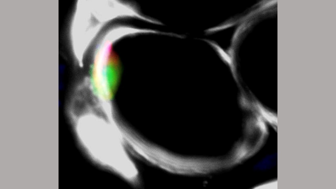
Image of mouse muscle. In white is mouse laminin, showing a cross section of muscle fibers. The green color marks a muscle stem cell protein called PAX7. The red color is for human-specific LaminA/C protein. Credit: Sunny Sun
In proof-of-concept experiments, Johns Hopkins Medicine scientists say they have successfully cultivated human muscle stem cells capable of renewing themselves and repairing muscle tissue damage in mice, potentially advancing efforts to treat muscle injuries and muscle-wasting disorders in people.
A report on the experiments was published April 7 in Cell Stem Cell.
To make the self-renewing stem cells, the scientists began with laboratory-grown human skin cells that were genetically reprogrammed to a more primitive state in which the cells have the potential to become almost any type of cell in the body. At this point, the cells are known as induced pluripotent stem (IPS) cells, and they are mixed with a solution of standard cell growth factors and nutrients that nudge them to differentiate into specific cell types.
In the laboratory, scientists have long been able to transform IPS cells into various types of cells, including skin and brain cells. What has been far more difficult, say the researchers, is the ability to turn IPS cells into self-renewing stem cells for a particular organ.
The research team, led by Gabsang Lee, Ph.D., D.V.M., professor of neurology and member of the Institute of Cell Engineering at Johns Hopkins Medicine, coaxed IPS cells to turn into muscle stem cells using a nutrient-rich broth. Further studies are planned, Lee says, to examine the recipe further to determine which ingredients may be key to brewing the muscle stem cells.
Lee is co-founder of Vita Therapeutics Inc., a Baltimore, Maryland-based cell engineering company, that hopes to bring muscle stem cell treatments to market for muscle wasting disorders, including muscular dystrophy. He cautions that such stem cell therapies are not yet available.
In proof-of-concept experiments with mice, the research team sought to determine where the newly developed cells would migrate in living animals, and if they could repair damaged tissue.
The team reported that when they injected the muscle stem cells into the mouse muscles, the cells moved to an area of the muscles known as the niche, where other natural muscle stem cells are typically found, and stayed there for more than four months.
The research team then used two different methods to determine if the muscle stem cells would repair damaged tissue.
In one method, the researchers transplanted the muscle stem cells into mice genetically engineered and bred without an immune system to avoid rejection of the transplanted cells. They then exposed the animals to a muscle-degrading toxin and radiation to eliminate muscle stem cells already existing within the mouse.
At the site of the toxin and radiation damage in the muscle tissue, the researchers found that the transplanted human muscle stem cells developed into myoblasts, a kind of muscle construction cell that repairs damage by fusing together and developing the microfibers that characterize normal muscle. They also found that some of the transplanted human muscle stem cells migrate to the niche and behave like muscle stem cells naturally found within the mouse.
In a second set of experiments, the researchers transplanted the muscle stem cells into mice genetically engineered with a mutation in the dystrophin gene, which results in Duchenne muscular dystrophy, a muscle wasting disorder in mice and humans.
The researchers found that transplanted muscle stem cells traveled to the muscle niche area. Over several months, tests showed the transplanted mice were able to run twice as far on mini treadmills than untreated mice, a measure of muscle strength.
"These muscle stem cells could potentially be developed as treatments for many types of muscle disorders," says Lee.
The research team plans to study the use of the cells in mouse models of other muscle-related conditions for their potential use in sports medicine, trauma, and age-related muscle loss.
Support for this research was provided by the National Institutes of Health (R01NS093213, R01-AR076390, K01-AR074048, R01AR070751), the Maryland Stem Cell Research Fund, the Maryland Stem Cell Fellowship, Vita Therapeutics, the Muscular Dystrophy Association, the Peter and Carmen Lucia Buck Foundation, the American Heart Association Predoctoral Fellowship, an American Heart Association Career Development Award, an American Heart Association Established Investigator Award, and National Research Foundation of Korea grants.
Under a license agreement between Vita Therapeutics and The Johns Hopkins University, Lee, Kathryn R. Wagner, and the University are entitled to royalty distributions related to technology described in the study discussed in this press release. Vita Therapeutics provided partial support for this study. Lee, Wagner, and Peter Andersen are co-founders of Vita Therapeutics and hold equity in the company.
Other scientists who contributed to the research include Sunny (Congshan) Sun, Suraj Kannan, In Young Choi, HoTae Lim, Hao Zhang, Grace Chen, Nancy Zhang, Seong-Hyun Park, Carlo Serra, Shama Iyer, Thomas Lloyd, Chulan Kwon and Peter Andersen of Johns Hopkins; Richard Lovering of the University of Maryland School of Medicine (now at the National Institutes of Health); Su Bin Lim of the Ajou University School of Medicine in South Korea; and Congshan Sun and Kathryn Wagner, formerly of Johns Hopkins and the Kennedy Krieger Institute and now at Vita Therapeutics and F. Hoffman La-Roche Inc., respectively.


