Scientists are exploring leading-edge technologies that could transform how cancer is studied, detected and treated by catching it earlier, when it's more treatable and survival rates are highest.
A new review by researchers at Oregon Health & Science University's Knight Cancer Institute and other universities highlights how advances in New Approach Methodologies and tissue engineering are offering powerful new tools to study the earliest stages of cancer development. New Approach Methodologies use human-relevant technologies such as in vitro tests, organoids, organs-on-a-chip and computational modeling to replace, reduce or refine animal testing.
These lab-grown models replicate the environment inside the human body and could unlock clues about how cancer begins.
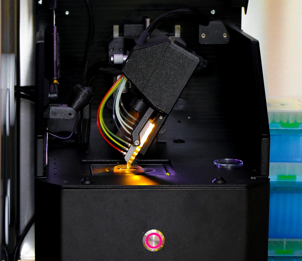
Nearly a decade after Luiz Bertassoni and his team earned national recognition for developing a groundbreaking method to 3D print blood vessels — a discovery named one of the year's top scientific breakthroughs by Discover magazine — Bertassoni is leveraging his earlier work to study complex cancers in ways traditional lab models can't. Now at the helm of a new chip-based system that more accurately mimics the human bone-tumor environment, Bertassoni and a cross-disciplinary team are using advanced bioengineering to create more realistic in-vitro models — a key step forward in the Food and Drug Administration's shift away from animal testing toward human-cell-based systems.

"Early detection is one of the most important factors in surviving cancer," said senior author Luiz Bertassoni, D.D.S., Ph.D., director of the Knight Cancer Precision Biofabrication Hub, and a professor at the OHSU Knight Cancer Institute and the OHSU School of Dentistry. "These new technologies give us a window into how cancer forms and progresses, which opens the door to understanding early cancer, paving the way for earlier diagnosis and even predict cancer initiation."
The review published today in the journal Nature Reviews Bioengineering.
Despite years of cancer research, scientists know relatively little about what happens in the body during the early stages of cancer. A major reason is the lack of access to early stage tumor samples, especially in organs that are hard to reach. Patients usually come to the clinic after symptoms are present, and that is usually too late.
Without samples of early cancer, it's difficult to understand the changes that occur as healthy tissue becomes cancerous.
Tissue engineering is helping close that gap. And recent technologies developed in the past decade, including many designed at the OHSU Knight Cancer Institute, have enabled scientists to mirror the complexity of cancer in the lab. These models, which have recently been prioritized as so-called New Approach Methodologies for medical research, let researchers precisely recreate and manipulate the early tumor environment, allowing them to test how specific cellular, genetic or environmental factors influence cancer development. This approach also supports the discovery of new biomarkers — biological red flags that could help clinicians detect cancer earlier and more accurately.
"This is a really exciting time in cancer research," Bertassoni said. "There is momentum in bringing together cancer biology, engineering and clinical treatment. There are so many avenues that didn't exist before."

Haylie Helms, M.S., an OHSU graduate student in biomedical engineering and a graduate fellow of the International Alliance for Cancer Early Detection, is lead author on the review.
Her dissertation is on engineering and biofabrication of early cancer models, paving the way for advanced cancer New Approach Methodologies. Specifically, she is researching single-cell 3D bioprinting as a tool for early cancer detection and treatment. Bioprinting allows for the creation of realistic, complex 3D tumor models that mimic the in vivo environment. These models can be used to study tumor development, responses to drugs and personalize treatment strategies.
"We can first build a healthy tissue and use different tools to turn it into cancer. We can also take live cancer cells from a patient biopsy and add them into the model," she said. "In the lab, we can watch and see, 'Why does a precancerous lesion in one person stay that way and never turn into cancer and in another person, it becomes a malignant tumor?'"
Their research emphasizes a growing focus on "cancer interception," intervening early, even before a tumor fully forms, to stop cancer in its tracks.
"A lot of the field is about late-stage cancer," Helms said. "We aim to understand and treat at the earliest possible moment."
In addition to Bertassoni and Helms, co-authors include Alexander Davies, D.V.M, Ph.D., Carolyn Schutt Ibsen, Ph.D., Ellen Langer, Ph.D., Rebekka Duhen, Ph.D., Sadik Esener, Ph.D., of OHSU; Prima Dewi Sinawang, Ph.D., Demir Akin, D.V.M., Ph.D., and Utkan Demirci, Ph.D., of Stanford University; and Rebecca C. Fitzgerald, M.D., of the University of Cambridge.
Research reported in this publication was supported by: the National Cancer Institute, the National Institute of General Medical Sciences, and the Office of the Director, all of the National Institutes of Health, under award numbers R21CA263860, R35GM151057, and K01OD031811; the National Science Foundation under award number 2339254; the OHSU Silver Family Innovation Award; ARCS Foundation Oregon; International Alliance for Cancer Early Detection, and the OHSU Cancer Early Detection Advanced Research Center. Ellen Langer, Ph.D. was supported by a Research Scholar Grant RSG-22-060-01-MM, from the American Cancer Society. The content is solely the responsibility of the authors and does not necessarily represent the official views of the NIH or other funders.


